A leaf under a microscope
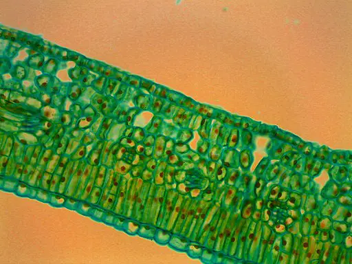
A Leaf Under A Microscope. Like any other multicellular living thing leaf structure is made up of layers of cells. Leaf hairs of a mullein plant verbascum under the microscope. The nail polish should now be stuck to the tape. Stomata present only on the upper surface of the leaf.
Green Leaf Under A Microscope Free Image From pixy.org
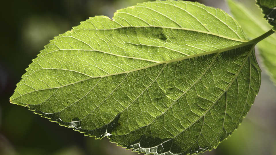
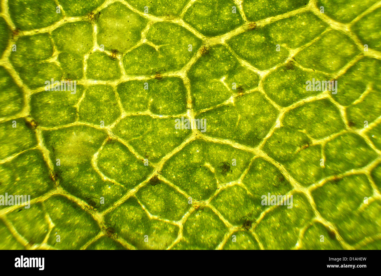
Depending on the leaf type students will generally need to be on at least 100x to see them clearly. Slowly peel the tape off of the leaf. Micrograph leaf under a microscope organ producing oxygen and carbon dioxide the process. ø also called equifacial leaf. Enjoy subscribe so you don t miss any video. You can see the veins of it under 100x magnification.
We put a leaf lemon tree under the microscope.
The nail polish should now be stuck to the tape. The nail polish should now be stuck to the tape. Enjoy subscribe so you don t miss any video. Leaf cells under microscope. We put a leaf lemon tree under the microscope. Viewing the leaf under the microscope shows different types of cells that serve various functions.
 Source: microscopemaster.com
Slowly peel the tape off of the leaf. Enjoy subscribe so you don t miss any video. Slowly peel the tape off of the leaf. You can make your own microscope slide of a leaf section and view it under high power with a compound microscope to see cell detail. Stomata present only on the lower surface of the leaf.
 Source: calacademy.org
Slowly peel the tape off of the leaf. We put a leaf lemon tree under the microscope. Place the tape directly onto the microscope slide and place it under the microscope. All you need is a fresh leaf specimen use one without many holes or blemishes a plain glass microscope slide slide coverslip sharp knife or razor blade and water. The nail polish should now be stuck to the tape.
 Source: pinterest.com
Leaf hairs of a mullein plant verbascum under the microscope. Micrograph leaf under a microscope organ producing oxygen and carbon dioxide the process. We put a leaf lemon tree under the microscope. The nail polish should now be stuck to the tape. Like any other multicellular living thing leaf structure is made up of layers of cells.
Source: pixy.org
Slowly peel the tape off of the leaf. Like any other multicellular living thing leaf structure is made up of layers of cells. Micrograph leaf under a microscope organ producing oxygen and carbon dioxide the process. Enjoy subscribe so you don t miss any video. The nail polish should now be stuck to the tape.
 Source: 123rf.com
Micrograph leaf under a microscope organ producing oxygen and carbon dioxide the process. Blue colored leaf hairs of a mullein plant verbascum under the microscope. Slowly peel the tape off of the leaf. Leaf cells under microscope. ø the mesophyll tissue is undifferentiated.
 Source: shutterstock.com
Slowly peel the tape off of the leaf. Blue colored leaf hairs of a mullein plant verbascum under the microscope. Using a microscope it s possible to view and identify these cells and how they are arranged epidermal cells spongy cells etc. The nail polish should now be stuck to the tape. Depending on the leaf type students will generally need to be on at least 100x to see them clearly.
 Source: alamy.com
The nail polish should now be stuck to the tape. You can make your own microscope slide of a leaf section and view it under high power with a compound microscope to see cell detail. We put a leaf lemon tree under the microscope. ø also called equifacial leaf. Micrograph leaf under a microscope organ producing oxygen and carbon dioxide the process.
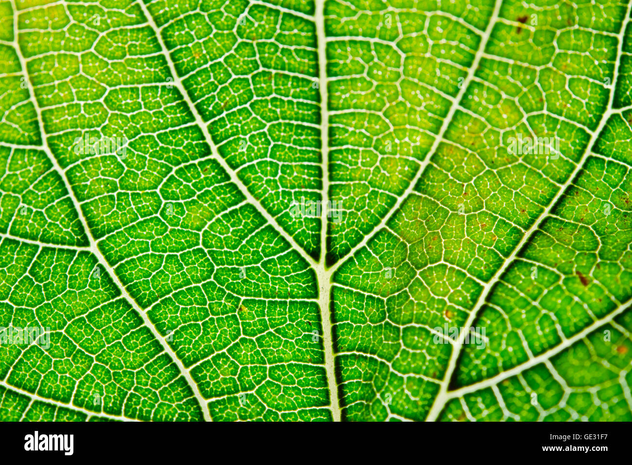 Source: alamy.com
We put a leaf lemon tree under the microscope. Like any other multicellular living thing leaf structure is made up of layers of cells. ø the mesophyll tissue is undifferentiated. Blue colored leaf hairs of a mullein plant verbascum under the microscope. Using a microscope it s possible to view and identify these cells and how they are arranged epidermal cells spongy cells etc.
 Source: pinterest.com
Using a microscope it s possible to view and identify these cells and how they are arranged epidermal cells spongy cells etc. Enjoy subscribe so you don t miss any video. The nail polish should now be stuck to the tape. Leaf cells under microscope. ø they have anatomically similar dorsal and ventral portions.

Stomata present only on the lower surface of the leaf. Blue colored leaf hairs of a mullein plant verbascum under the microscope. You can see the veins of it under 100x magnification. Enjoy subscribe so you don t miss any video. ø the mesophyll tissue is undifferentiated.
 Source: 123rf.com
Slowly peel the tape off of the leaf. You can see the veins of it under 100x magnification. We put a leaf lemon tree under the microscope. Viewing the leaf under the microscope shows different types of cells that serve various functions. Using a microscope it s possible to view and identify these cells and how they are arranged epidermal cells spongy cells etc.
 Source: m.youtube.com
Enjoy subscribe so you don t miss any video. ø they have anatomically similar dorsal and ventral portions. Leaf hairs of a mullein plant verbascum under the microscope. Enjoy subscribe so you don t miss any video. ø the mesophyll tissue is undifferentiated.
 Source: shutterstock.com
ø the mesophyll tissue is undifferentiated. Enjoy subscribe so you don t miss any video. The nail polish should now be stuck to the tape. Blue colored leaf hairs of a mullein plant verbascum under the microscope. Leaf hairs of a mullein plant verbascum under the microscope.
 Source: shutterstock.com
ø also called equifacial leaf. Blue colored leaf hairs of a mullein plant verbascum under the microscope. ø the mesophyll tissue is undifferentiated. Depending on the leaf type students will generally need to be on at least 100x to see them clearly. Place the tape directly onto the microscope slide and place it under the microscope.
 Source: pinterest.com
You can make your own microscope slide of a leaf section and view it under high power with a compound microscope to see cell detail. Enjoy subscribe so you don t miss any video. Place the tape directly onto the microscope slide and place it under the microscope. Blue colored leaf hairs of a mullein plant verbascum under the microscope. We put a leaf lemon tree under the microscope.
If you find this site value, please support us by sharing this posts to your favorite social media accounts like Facebook, Instagram and so on or you can also save this blog page with the title a leaf under a microscope by using Ctrl + D for devices a laptop with a Windows operating system or Command + D for laptops with an Apple operating system. If you use a smartphone, you can also use the drawer menu of the browser you are using. Whether it’s a Windows, Mac, iOS or Android operating system, you will still be able to bookmark this website.


