Abdominal cavity diagram
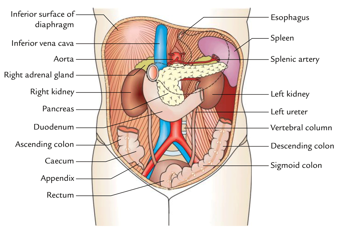
Abdominal Cavity Diagram. We are pleased to provide you with the picture named abdomen arteries veins and duct diagram we hope this picture abdomen arteries veins and duct diagram can help you study and research. It follows the thorax or cephalothorax. Ultrasound can detect problems in most abdominal organs. Connective tissue called the mesentery holds the abdominal organs together.
 Image Result For Organs Abdominal Cavity Body Organs Diagram Human Organ Diagram Human Body Diagram From pinterest.com
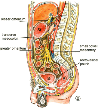
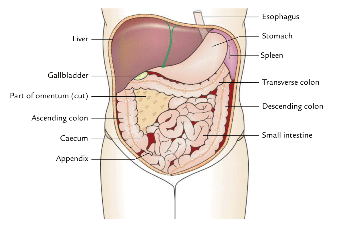
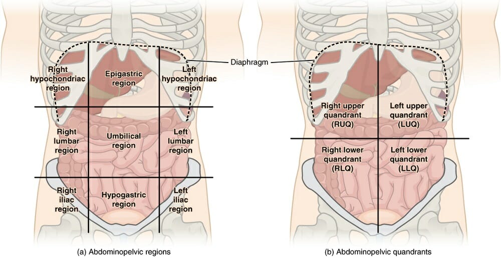
The abdominal cavity is a large cavity found in the torso of mammals between the thoracic cavity which it is separated from by the thoracic diaphragm and the pelvic cavity a protective layer that is called the peritoneum which plays a role in immunity supporting organs and fat storage lines the abdominal cavity. The liver is located in the upper right hand part of the abdominal cavity under the ribs. The abdomen colloquially called the stomach belly tummy or midriff is the part of the body between the thorax chest and pelvis in humans and in other vertebrates the abdomen is the front part of the abdominal segment of the trunk the area occupied by the abdomen is called the abdominal cavity in arthropods it is the posterior tagma of the body. Its lower boundary is the upper plane of the pelvic cavity. The abdomen consists of. Ultrasound can detect problems in most abdominal organs.
The abdomen consists of.
Its upper boundary is the diaphragm a sheet of muscle and connective tissue that separates it from the chest cavity. The abdominal cavity is a large cavity found in the torso of mammals between the thoracic cavity which it is separated from by the thoracic diaphragm and the pelvic cavity a protective layer that is called the peritoneum which plays a role in immunity supporting organs and fat storage lines the abdominal cavity. The abdomen colloquially called the stomach belly tummy or midriff is the part of the body between the thorax chest and pelvis in humans and in other vertebrates the abdomen is the front part of the abdominal segment of the trunk the area occupied by the abdomen is called the abdominal cavity in arthropods it is the posterior tagma of the body. The inguinal region contains the inguinal canal that carries the spermatic cord in men and the round ligaments in women. It also gives structural support to the upper body. We are pleased to provide you with the picture named abdomen arteries veins and duct diagram we hope this picture abdomen arteries veins and duct diagram can help you study and research.
 Source: radiologykey.com
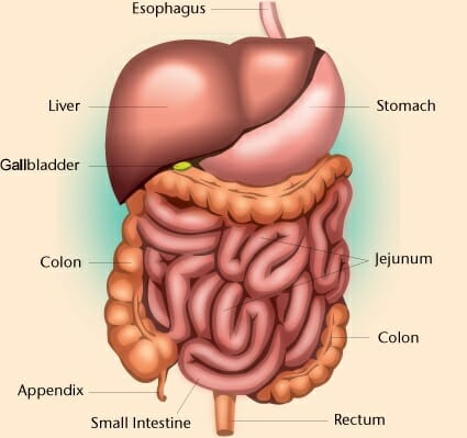
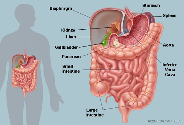
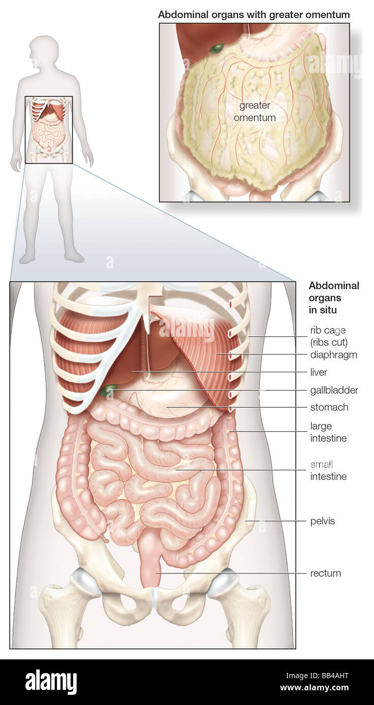
It is a flexible dynamic container housing most of the organs of the alimentary system and part of the urogenital system. The liver is located in the upper right hand part of the abdominal cavity under the ribs. It follows the thorax or cephalothorax. Abdominal cavity largest hollow space of the body. These general diagrams show the digestive system with the major human anatomical structures labeled mouth tongue oral cavity teeth buccal glands throat pharynx oesophagus stomach small intestine large intestine liver gall bladder and pancreas.
 Source: earthslab.com
Vertically it is enclosed by the vertebral column and the abdominal and other muscles. It also gives structural support to the upper body. A probe on the abdomen reflects high frequency sound waves off the abdominal organs creating images on a screen. The abdomen consists of. Although it has many functions the liver is best known for processing blood separating waste from.
 Source: biologydictionary.net
The abdomen consists of. For more anatomy content please follow us and visit our website. Peritoneum serves as the lining of the abdominal cavity. It also gives structural support to the upper body. The abdominal cavity is a large cavity found in the torso of mammals between the thoracic cavity which it is separated from by the thoracic diaphragm and the pelvic cavity a protective layer that is called the peritoneum which plays a role in immunity supporting organs and fat storage lines the abdominal cavity.
 Source: quizlet.com
Connective tissue called the mesentery holds the abdominal organs together. It follows the thorax or cephalothorax. These general diagrams show the digestive system with the major human anatomical structures labeled mouth tongue oral cavity teeth buccal glands throat pharynx oesophagus stomach small intestine large intestine liver gall bladder and pancreas. It also gives structural support to the upper body. The abdomen colloquially called the stomach belly tummy or midriff is the part of the body between the thorax chest and pelvis in humans and in other vertebrates the abdomen is the front part of the abdominal segment of the trunk the area occupied by the abdomen is called the abdominal cavity in arthropods it is the posterior tagma of the body.
Source: quora.com
Peritoneum serves as the lining of the abdominal cavity. Peritoneum serves as the lining of the abdominal cavity. The abdomen colloquially called the stomach belly tummy or midriff is the part of the body between the thorax chest and pelvis in humans and in other vertebrates the abdomen is the front part of the abdominal segment of the trunk the area occupied by the abdomen is called the abdominal cavity in arthropods it is the posterior tagma of the body. It follows the thorax or cephalothorax. Its upper boundary is the diaphragm a sheet of muscle and connective tissue that separates it from the chest cavity.
 Source: emsworld.com
Full labeled anatomical diagrams anatomy of the abdomen and digestive system. The abdomen consists of. Although it has many functions the liver is best known for processing blood separating waste from. Abdomen the abdomen is the part of the trunk between the thorax and the pelvis. The function of the anterior abdominal wall is to keep and protect the viscera lining the abdomen.
 Source: biologydictionary.net
Connective tissue called the mesentery holds the abdominal organs together. Connective tissue called the mesentery holds the abdominal organs together. The inguinal region contains the inguinal canal that carries the spermatic cord in men and the round ligaments in women. Ultrasound can detect problems in most abdominal organs. The abdominal cavity is a large cavity found in the torso of mammals between the thoracic cavity which it is separated from by the thoracic diaphragm and the pelvic cavity a protective layer that is called the peritoneum which plays a role in immunity supporting organs and fat storage lines the abdominal cavity.
 Source: pinterest.com
Peritoneum serves as the lining of the abdominal cavity. We are pleased to provide you with the picture named abdomen arteries veins and duct diagram we hope this picture abdomen arteries veins and duct diagram can help you study and research. Its upper boundary is the diaphragm a sheet of muscle and connective tissue that separates it from the chest cavity. Although it has many functions the liver is best known for processing blood separating waste from. Ultrasound can detect problems in most abdominal organs.
 Source: earthslab.com
Its upper boundary is the diaphragm a sheet of muscle and connective tissue that separates it from the chest cavity. Peritoneum serves as the lining of the abdominal cavity. The liver is located in the upper right hand part of the abdominal cavity under the ribs. Although it has many functions the liver is best known for processing blood separating waste from. The abdominal cavity is a large cavity found in the torso of mammals between the thoracic cavity which it is separated from by the thoracic diaphragm and the pelvic cavity a protective layer that is called the peritoneum which plays a role in immunity supporting organs and fat storage lines the abdominal cavity.
 Source: britannica.com
The inguinal region contains the inguinal canal that carries the spermatic cord in men and the round ligaments in women. The liver is located in the upper right hand part of the abdominal cavity under the ribs. Ultrasound can detect problems in most abdominal organs. It is a flexible dynamic container housing most of the organs of the alimentary system and part of the urogenital system. Abdominal cavity largest hollow space of the body.
 Source: en.wikipedia.org
Abdominal cavity largest hollow space of the body. Its lower boundary is the upper plane of the pelvic cavity. Connective tissue called the mesentery holds the abdominal organs together. Abdominal cavity largest hollow space of the body. A probe on the abdomen reflects high frequency sound waves off the abdominal organs creating images on a screen.
 Source: webmd.com
These general diagrams show the digestive system with the major human anatomical structures labeled mouth tongue oral cavity teeth buccal glands throat pharynx oesophagus stomach small intestine large intestine liver gall bladder and pancreas. The liver is located in the upper right hand part of the abdominal cavity under the ribs. The inguinal region contains the inguinal canal that carries the spermatic cord in men and the round ligaments in women. The abdominal cavity is a large cavity found in the torso of mammals between the thoracic cavity which it is separated from by the thoracic diaphragm and the pelvic cavity a protective layer that is called the peritoneum which plays a role in immunity supporting organs and fat storage lines the abdominal cavity. We are pleased to provide you with the picture named abdomen arteries veins and duct diagram we hope this picture abdomen arteries veins and duct diagram can help you study and research.
 Source: alamy.com
Connective tissue called the mesentery holds the abdominal organs together. Ultrasound can detect problems in most abdominal organs. Peritoneum serves as the lining of the abdominal cavity. The inguinal region contains the inguinal canal that carries the spermatic cord in men and the round ligaments in women. Its lower boundary is the upper plane of the pelvic cavity.
 Source: pinterest.com
Abdominal walls abdominal cavity abdominal viscera. A probe on the abdomen reflects high frequency sound waves off the abdominal organs creating images on a screen. The abdominal cavity is a large cavity found in the torso of mammals between the thoracic cavity which it is separated from by the thoracic diaphragm and the pelvic cavity a protective layer that is called the peritoneum which plays a role in immunity supporting organs and fat storage lines the abdominal cavity. Although it has many functions the liver is best known for processing blood separating waste from. Its lower boundary is the upper plane of the pelvic cavity.
 Source: pinterest.com
These general diagrams show the digestive system with the major human anatomical structures labeled mouth tongue oral cavity teeth buccal glands throat pharynx oesophagus stomach small intestine large intestine liver gall bladder and pancreas. Full labeled anatomical diagrams anatomy of the abdomen and digestive system. Its upper boundary is the diaphragm a sheet of muscle and connective tissue that separates it from the chest cavity. These general diagrams show the digestive system with the major human anatomical structures labeled mouth tongue oral cavity teeth buccal glands throat pharynx oesophagus stomach small intestine large intestine liver gall bladder and pancreas. The abdomen consists of.
If you find this site beneficial, please support us by sharing this posts to your own social media accounts like Facebook, Instagram and so on or you can also bookmark this blog page with the title abdominal cavity diagram by using Ctrl + D for devices a laptop with a Windows operating system or Command + D for laptops with an Apple operating system. If you use a smartphone, you can also use the drawer menu of the browser you are using. Whether it’s a Windows, Mac, iOS or Android operating system, you will still be able to bookmark this website.


