Allium root tip labeled
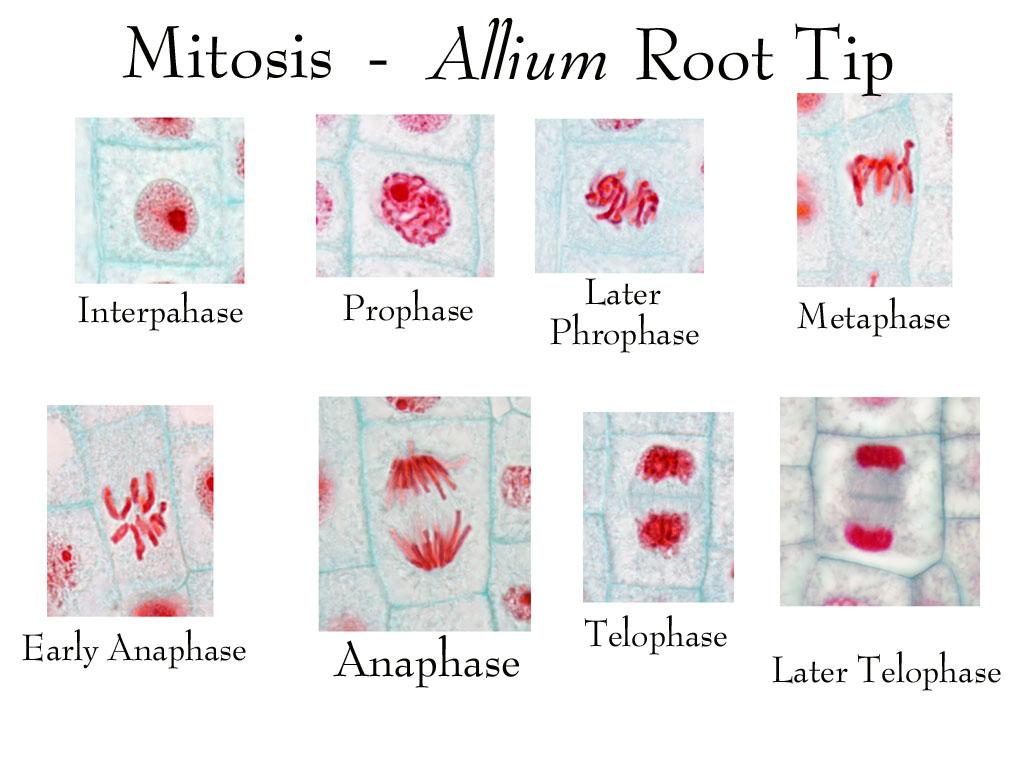
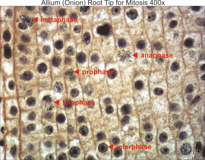
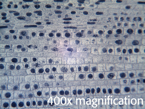
Allium Root Tip Labeled. Telophase the final phase of cell division will appear as two nuclei are formed and have. Ages 8 in stock ready to ship need it fast. If you have a microscope 400x and a properly stained slide of the onion root tip or allium root tip you can see the phases in different cells frozen in time. The allium roots need to be prepared 1 10 days in advance of the lesson.
 Cell Cycle And Mitosis Laboratory Notes For Bio 1003 From faculty.baruch.cuny.edu
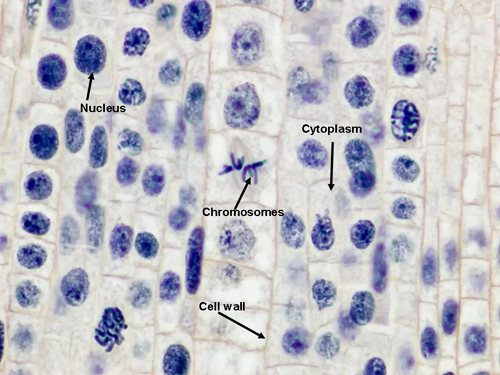
1 the allium root tip slide was obtained and placed on the stage of the microscope. You will be looking at strands of dna inside the cell. These cells are not undergoing mitosis. Onion root mitosis allium root tip. Ages 8 in stock ready to ship need it fast. The chromosomes are easily observed through a compound light microscope.
Examine the square cells just inside the root cap.
These cells are not undergoing mitosis. A monocot root tip with all stages of mitosis visible. It is common to see photomicrographs of onion root cells when demonstrating how cell division takes place in plants. See delivery options in cart. The onion root tip slide is included free in your slide kit when you purchase a microscope from microscope world. Light micrograph of onion allium cepa root tip cells stained with acetocarmine to show nuclei and chromosomes.

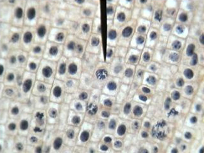
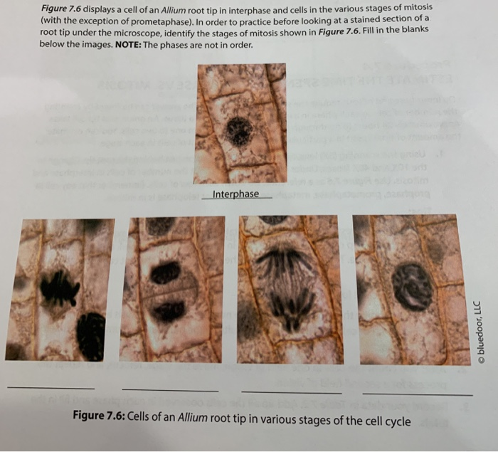
Obtain a prepared slide of an onion allium root tip. Ages 8 in stock ready to ship need it fast. The field includes cells in interphase prophase metaphase and late telophase. Telophase the final phase of cell division will appear as two nuclei are formed and have. Examine the square cells just inside the root cap.
 Source: search.library.wisc.edu
If you have a microscope 400x and a properly stained slide of the onion root tip or allium root tip you can see the phases in different cells frozen in time. The root tip the root apical meristem e1. Allium onion root tip slide l s. You will be looking at strands of dna inside the cell. Some practitioners report that cutting the root tips around noon makes a difference to the mitotic index so you may want your technician to cut and fix the tips in ethanoic alcohol rather than ask your students to carry out this step.
 Source: sciencegeek.net
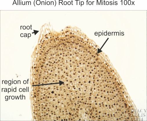
Telophase the final phase of cell division will appear as two nuclei are formed and have. This slide is a longitudinal section labeled as l s. The chromosomes are easily observed through a compound light microscope. The centre root tip meristem was focused under medium power. This is the root meristem embryonic tissue where mitosis is occurring.
 Source: chegg.com
A monocot root tip with all stages of mitosis visible. Ages 8 in stock ready to ship need it fast. The field includes cells in interphase prophase metaphase and late telophase. Light micrograph of onion allium cepa root tip cells stained with acetocarmine to show nuclei and chromosomes. These cells are not undergoing mitosis.
 Source: www-plb.ucdavis.edu
Allium onion root tip slide l s. Telophase the final phase of cell division will appear as two nuclei are formed and have. Talk to an expert. Discover and save your own pins on pinterest. The field includes cells in interphase prophase metaphase and late telophase.
 Source: dissectionconnection.com.au
The field includes cells in interphase prophase metaphase and late telophase. Feb 23 2015 this pin was discovered by kelsey robbins. Telophase the final phase of cell division will appear as two nuclei are formed and have. This is the root meristem embryonic tissue where mitosis is occurring. Light micrograph of onion allium cepa root tip cells stained with acetocarmine to show nuclei and chromosomes.
 Source: pinterest.com
Examine the cells along the root tip from the natural terminal end rounded to the cut end. These cells are not undergoing mitosis. Examine the cells along the root tip from the natural terminal end rounded to the cut end. If you have a microscope 400x and a properly stained slide of the onion root tip or allium root tip you can see the phases in different cells frozen in time. Discover and save your own pins on pinterest.
 Source: dissectionconnection.com.au
Farther up the root is the elongation zone where cells are long rectangles. Light micrograph of onion allium cepa root tip cells stained with acetocarmine to show nuclei and chromosomes. 2 a proper biological diagram of the meristem was drawn. Microscope allium root tip slide method. Examine the square cells just inside the root cap.
 Source: homesciencetools.com
Light micrograph of onion allium cepa root tip cells stained with acetocarmine to show nuclei and chromosomes. Onions have larger chromosomes than most plants and stain dark. See delivery options in cart. Ages 8 in stock ready to ship need it fast. Allium onion root tip slide l s.
 Source: semanticscholar.org
It is common to see photomicrographs of onion root cells when demonstrating how cell division takes place in plants. The root tip the root apical meristem e1. Allium onion root tip slide l s. Telophase the final phase of cell division will appear as two nuclei are formed and have. The field includes cells in interphase prophase metaphase and late telophase.

1 the allium root tip slide was obtained and placed on the stage of the microscope. The root apical meristem is at the tip behind the root cap and consists of. The allium roots need to be prepared 1 10 days in advance of the lesson. You will be looking at strands of dna inside the cell. Some practitioners report that cutting the root tips around noon makes a difference to the mitotic index so you may want your technician to cut and fix the tips in ethanoic alcohol rather than ask your students to carry out this step.
 Source: quizlet.com
If you have a microscope 400x and a properly stained slide of the onion root tip or allium root tip you can see the phases in different cells frozen in time. Onion root mitosis allium root tip. Onions have larger chromosomes than most plants and stain dark. Feb 23 2015 this pin was discovered by kelsey robbins. 2 a proper biological diagram of the meristem was drawn.

1 the allium root tip slide was obtained and placed on the stage of the microscope. The onion root tip slide is included free in your slide kit when you purchase a microscope from microscope world. 2 a proper biological diagram of the meristem was drawn. This is the root meristem embryonic tissue where mitosis is occurring. If you have a microscope 400x and a properly stained slide of the onion root tip or allium root tip you can see the phases in different cells frozen in time.
 Source: chegg.com
Ages 8 in stock ready to ship need it fast. Examine the cells along the root tip from the natural terminal end rounded to the cut end. Onion root mitosis allium root tip. Talk to an expert. 1 the allium root tip slide was obtained and placed on the stage of the microscope.
 Source: faculty.baruch.cuny.edu
The root apical meristem is at the tip behind the root cap and consists of. You will be looking at strands of dna inside the cell. Feb 23 2015 this pin was discovered by kelsey robbins. Onion root mitosis allium root tip. Telophase the final phase of cell division will appear as two nuclei are formed and have.
If you find this site convienient, please support us by sharing this posts to your favorite social media accounts like Facebook, Instagram and so on or you can also bookmark this blog page with the title allium root tip labeled by using Ctrl + D for devices a laptop with a Windows operating system or Command + D for laptops with an Apple operating system. If you use a smartphone, you can also use the drawer menu of the browser you are using. Whether it’s a Windows, Mac, iOS or Android operating system, you will still be able to bookmark this website.


