Amoeba proteus under microscope
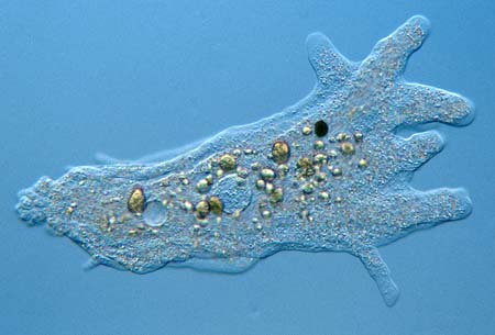
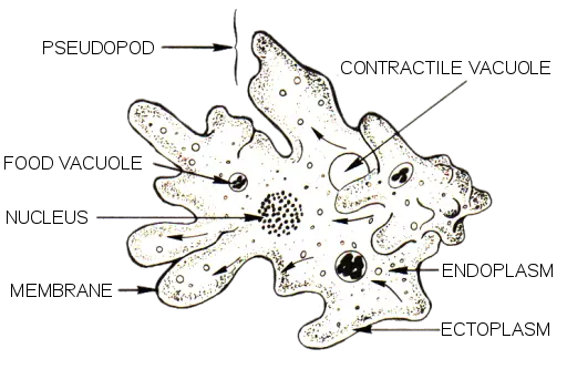
Amoeba Proteus Under Microscope. These microvilli can help amoeba proteus attach and release from the surface of the substrate. Amoeba under the microscope fixing staining techniques and structure amoeba plural amoebas amoebae is a genus that belongs to kingdom protozoa. It can almost be seen with the naked eye. An amoeba s cell s organelles and cytoplasm are enclosed by the membrane.
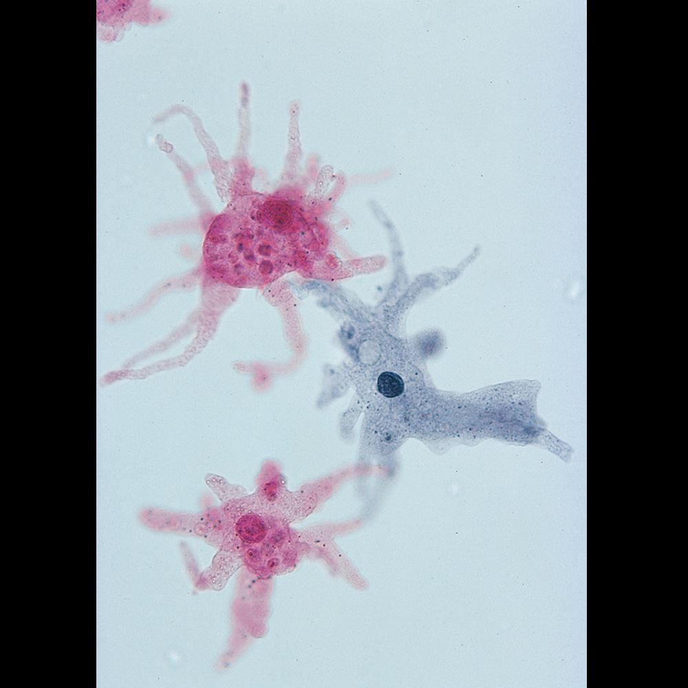 Amoeba Proteus Slide W M Microscope Sample Slides Amazon Com Industrial Scientific From amazon.com
In fact the outside face of the membrane has many microvilli attached to it can only be seen under an electron microscope. Image of amoeba captured with the digital ba210 microscope at 100x magnification. Amoeba under the microscope fixing staining techniques and structure amoeba plural amoebas amoebae is a genus that belongs to kingdom protozoa. Generally the term is used to describe single celled organisms that move in a primitive crawling manner by using temporary false feet known as pseudopods. Image courtesy pearson scott foresman. These microvilli can help amoeba proteus attach and release from the surface of the substrate.
In fact the outside face of the membrane has many microvilli attached to it can only be seen under an electron microscope.
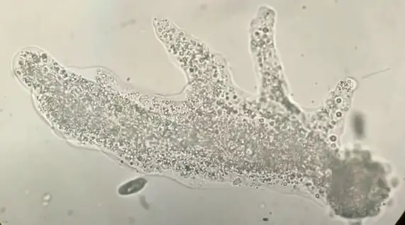
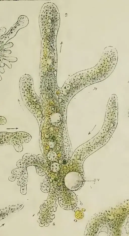
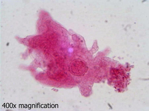
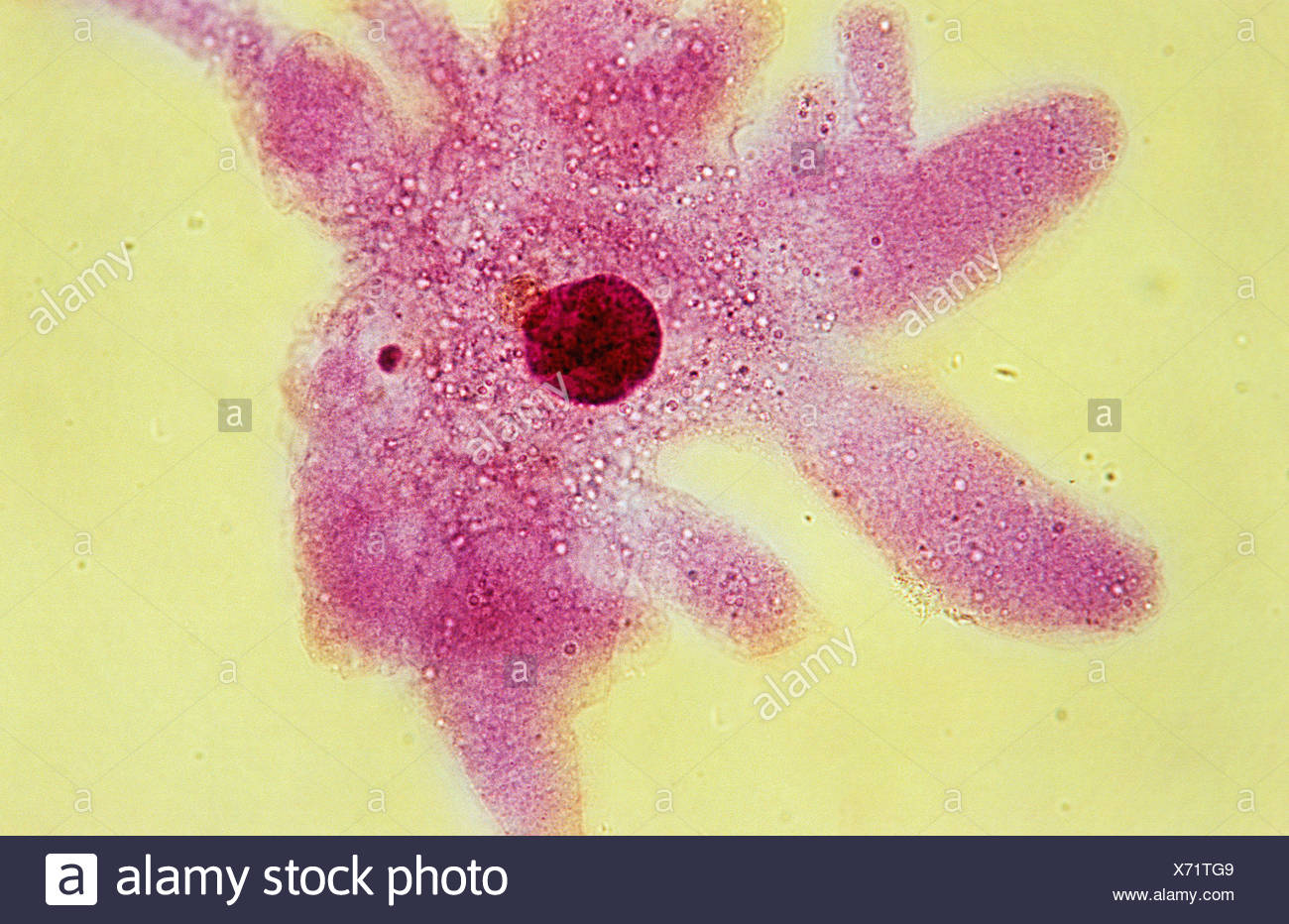
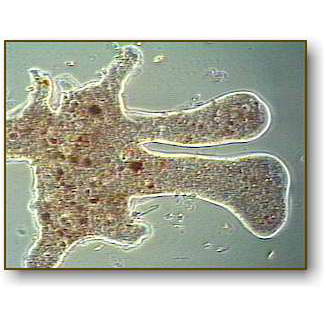
Amoebas are usually considered among the lowest and most primitive forms of life. In fact the outside face of the membrane has many microvilli attached to it can only be seen under an electron microscope. Amoeba are shapeless they look like a big blob unicellular organisms from the genus protozoa. Protozoa in this group move and gather food using pseudopodia or false feet as can be seen on many specimens in this slide. Amoeba proteus the real life microscopic blob amoeba proteus is a microscopic protist found in lakes and ponds that moves by stretching its pseudopodia fal. Consequently what magnification do you need to see amoeba.
 Source: microscopeclarity.com
Amoeba proteus slide w m microscope sample slides amazon com. An amoeba s cell s organelles and cytoplasm are enclosed by the membrane. It has an ever changing shape and is approximately 500 1000µnm long. Lab manual exercise 1. Use this whole mount microscope slide to study the basic characteristics of the popular protist amoeba.
 Source: m.youtube.com
Amoeba are shapeless they look like a big blob unicellular organisms from the genus protozoa. An amoeba s cell s organelles and cytoplasm are enclosed by the membrane. Amoeba are shapeless they look like a big blob unicellular organisms from the genus protozoa. Image of amoeba captured with the digital ba210 microscope at 100x magnification. It can almost be seen with the naked eye.
 Source: amazon.com
It has an ever changing shape and is approximately 500 1000µnm long. The third secret of amoeba proteus is its cell membrane is not that smooth like it shows under the optical microscope. An amoeba s cell s organelles and cytoplasm are enclosed by the membrane. A tiny blob of colorless jelly with a dark speck inside it this is what an amoeba looks like when seen through a microscope. Amoeba proteus is a single celled organism that belongs to the phylum sarcodina.
 Source: microscopemaster.com
Image of amoeba captured with the digital ba210 microscope at 100x magnification. Lab manual exercise 1. Protozoa in this group move and gather food using pseudopodia or false feet as can be seen on many specimens in this slide. Amoeba under the microscope fixing staining techniques and structure amoeba plural amoebas amoebae is a genus that belongs to kingdom protozoa. It can almost be seen with the naked eye.
 Source: britannica.com
An amoeba s cell s organelles and cytoplasm are enclosed by the membrane. Bio1140 lab 3 cellular processes in amoeba proteus ppt video. These microvilli can help amoeba proteus attach and release from the surface of the substrate. Lab manual exercise 1. Protozoa in this group move and gather food using pseudopodia or false feet as can be seen on many specimens in this slide.
 Source: youtube.com
Amoeba proteus is a single celled organism that belongs to the phylum sarcodina. An amoeba s cell s organelles and cytoplasm are enclosed by the membrane. Bio1140 lab 3 cellular processes in amoeba proteus ppt video. Amoeba proteus is a single celled organism that belongs to the phylum sarcodina. Sarcodina amoeba proteus microscope slide durable modeling.
 Source: homesciencetools.com
Amoeba under the microscope fixing staining techniques and structure amoeba plural amoebas amoebae is a genus that belongs to kingdom protozoa. Other species of amoebas are either too small too fragile or atypical in structure. Consequently what magnification do you need to see amoeba. It can almost be seen with the naked eye. Image of amoeba captured with the digital ba210 microscope at 100x magnification.
 Source: microscopy-uk.org.uk
Amoeba under the microscope fixing staining techniques and structure amoeba plural amoebas amoebae is a genus that belongs to kingdom protozoa. Amoeba proteus slide w m microscope sample slides amazon com. Generally the term is used to describe single celled organisms that move in a primitive crawling manner by using temporary false feet known as pseudopods. Use this whole mount microscope slide to study the basic characteristics of the popular protist amoeba. Other species of amoebas are either too small too fragile or atypical in structure.
 Source: rsscience.com
Generally the term is used to describe single celled organisms that move in a primitive crawling manner by using temporary false feet known as pseudopods. Lab manual exercise 1. Amoeba proteus is a single celled organism that belongs to the phylum sarcodina. Amoeba proteus slide w m microscope sample slides amazon com. It has an ever changing shape and is approximately 500 1000µnm long.
 Source: youtube.com
Image of amoeba captured with the digital ba210 microscope at 100x magnification. Amoeba proteus is a single celled organism that belongs to the phylum sarcodina. Generally the term is used to describe single celled organisms that move in a primitive crawling manner by using temporary false feet known as pseudopods. Protozoa in this group move and gather food using pseudopodia or false feet as can be seen on many specimens in this slide. The amoeba proteus is a large protozoan and belongs to the phyllum sarcodina.
 Source: alamy.com
Amoeba proteus is a single celled organism that belongs to the phylum sarcodina. The amoeba proteus can be ordered from science supply companies and is the classic specimen used in the classroom to demonstrate the pseudopods in action. An amoeba s cell s organelles and cytoplasm are enclosed by the membrane. Amoeba proteus slide w m microscope sample slides amazon com. Protozoa in this group move and gather food using pseudopodia or false feet as can be seen on many specimens in this slide.
 Source: davidwangblog.wordpress.com
Consequently what magnification do you need to see amoeba. An amoeba s cell s organelles and cytoplasm are enclosed by the membrane. Image of amoeba captured with the digital ba210 microscope at 100x magnification. It can almost be seen with the naked eye. The amoeba proteus can be ordered from science supply companies and is the classic specimen used in the classroom to demonstrate the pseudopods in action.
 Source: pinterest.com
Sarcodina amoeba proteus microscope slide durable modeling. Amoeba under the microscope fixing staining techniques structure. In fact the outside face of the membrane has many microvilli attached to it can only be seen under an electron microscope. Consequently what magnification do you need to see amoeba. An amoeba s cell s organelles and cytoplasm are enclosed by the membrane.
 Source: microscopemaster.com
Amoeba proteus is a single celled organism that belongs to the phylum sarcodina. Image courtesy pearson scott foresman. Amoeba proteus the real life microscopic blob amoeba proteus is a microscopic protist found in lakes and ponds that moves by stretching its pseudopodia fal. The amoeba proteus can be ordered from science supply companies and is the classic specimen used in the classroom to demonstrate the pseudopods in action. Lab manual exercise 1.
 Source: microscope-microscope.org
Image of amoeba captured with the digital ba210 microscope at 100x magnification. The third secret of amoeba proteus is its cell membrane is not that smooth like it shows under the optical microscope. These microvilli can help amoeba proteus attach and release from the surface of the substrate. Bio1140 lab 3 cellular processes in amoeba proteus ppt video. Amoeba proteus slide w m microscope sample slides amazon com.
If you find this site helpful, please support us by sharing this posts to your favorite social media accounts like Facebook, Instagram and so on or you can also save this blog page with the title amoeba proteus under microscope by using Ctrl + D for devices a laptop with a Windows operating system or Command + D for laptops with an Apple operating system. If you use a smartphone, you can also use the drawer menu of the browser you are using. Whether it’s a Windows, Mac, iOS or Android operating system, you will still be able to bookmark this website.


