Anatomy of a cow eye
Anatomy Of A Cow Eye. Thus the first job of the dissector is to remove the connective tissue to produce a neatly trimmed eyeball leaving the eyeball itself and the rear cord intact. Rods respond in dim light. The place where the optic nerve leaves the retina. When a calf is born both parts of the skull are almost the same size.
 Solved 1 Identify The Labeled Structures In The Accompan Chegg Com From chegg.com
Thus the first job of the dissector is to remove the connective tissue to produce a neatly trimmed eyeball leaving the eyeball itself and the rear cord intact. Each eye has a blind spot where there are no light sensitive cells. The iris is the part of the eye that is colored. Anatomy of a cow eye slideshare uses cookies to improve functionality and performance and to provide you with relevant advertising. Rods respond in dim light. About press copyright contact us creators advertise developers terms privacy policy safety how youtube works test new features press copyright contact us creators.
If you continue browsing the site you agree to the use of cookies on this website.
Each eye has a blind spot where there are no light sensitive cells. Rods respond in dim light. The front is responsible for the front part of the face of the cow. A tough clear covering over the iris and pupil that protects t. Thus the first job of the dissector is to remove the connective tissue to produce a neatly trimmed eyeball leaving the eyeball itself and the rear cord intact. This area is covered in many colors.
 Source: homesciencetools.com
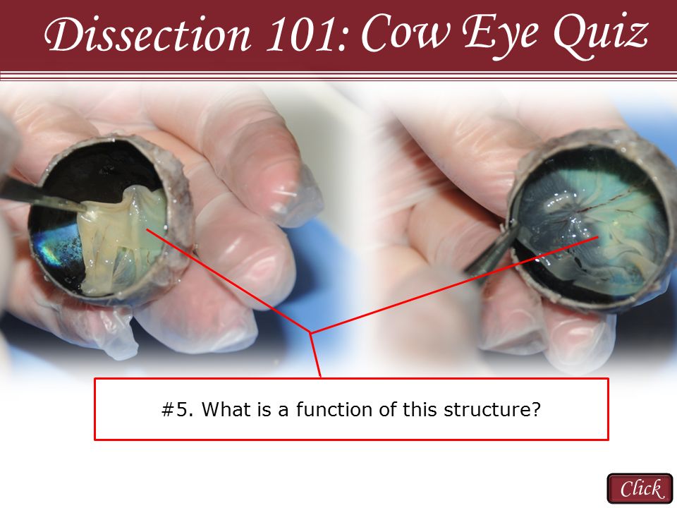
This area is covered in many colors. If you continue browsing the site you agree to the use of cookies on this website. When a calf is born both parts of the skull are almost the same size. However with age the facial part becomes larger and in size begins to prevail over the brain. A thick jelly that helps give the eyeball its shape.
 Source: slideshare.net
The front is responsible for the front part of the face of the cow. A cow s eye as normally provided will not be a neatly trimmed eyeball but a messy agglomeration of flesh. This area is covered in many colors. Each eye has a blind spot where there are no light sensitive cells. The iris has muscles around the pupil to help it to expand in bright light and to contract in dim light.
 Source: pinterest.com
A thick jelly that helps give the eyeball its shape. About press copyright contact us creators advertise developers terms privacy policy safety how youtube works test new features press copyright contact us creators. The iris is the part of the eye that is colored. Each eye has a blind spot where there are no light sensitive cells. When a calf is born both parts of the skull are almost the same size.
 Source: biologyjunction.com
A dark hole in the center of the iris that lets light into the. The iris has muscles around the pupil to help it to expand in bright light and to contract in dim light. About press copyright contact us creators advertise developers terms privacy policy safety how youtube works test new features press copyright contact us creators. Thus the first job of the dissector is to remove the connective tissue to produce a neatly trimmed eyeball leaving the eyeball itself and the rear cord intact. About press copyright contact us creators advertise developers terms privacy policy safety how youtube works test new features press copyright contact us creators.
 Source: prezi.com
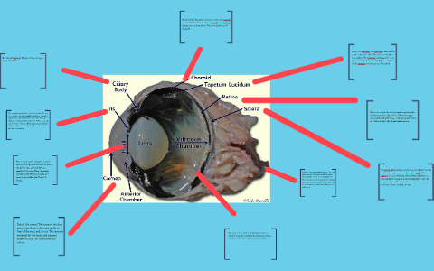
A cow s eye as normally provided will not be a neatly trimmed eyeball but a messy agglomeration of flesh. About press copyright contact us creators advertise developers terms privacy policy safety how youtube works test new features press copyright contact us creators. These include eye sockets nasal cavity and mouth. The tapetum can be used for cows to see in the dark because the light can reflect on the tapetum so they can see in the dark. Thus the first job of the dissector is to remove the connective tissue to produce a neatly trimmed eyeball leaving the eyeball itself and the rear cord intact.
 Source: fr.pinterest.com
Each eye has a blind spot where there are no light sensitive cells. However with age the facial part becomes larger and in size begins to prevail over the brain. If you continue browsing the site you agree to the use of cookies on this website. The place where the optic nerve leaves the retina. These include eye sockets nasal cavity and mouth.
 Source: pinterest.com
However with age the facial part becomes larger and in size begins to prevail over the brain. Rods respond in dim light. The front is responsible for the front part of the face of the cow. This area is covered in many colors. A cow s eye as normally provided will not be a neatly trimmed eyeball but a messy agglomeration of flesh.
 Source: slideplayer.com
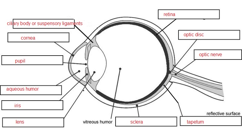
A tough clear covering over the iris and pupil that protects t. The place where the optic nerve leaves the retina. The iris is the part of the eye that is colored. The iris has muscles around the pupil to help it to expand in bright light and to contract in dim light. A tough clear covering over the iris and pupil that protects t.
 Source: quizlet.com
When a calf is born both parts of the skull are almost the same size. Thus the first job of the dissector is to remove the connective tissue to produce a neatly trimmed eyeball leaving the eyeball itself and the rear cord intact. About press copyright contact us creators advertise developers terms privacy policy safety how youtube works test new features press copyright contact us creators. The bundle of nerve fibers that carry information from the retina to the brain. This area is covered in many colors.
 Source: biologycorner.com
This area is covered in many colors. The iris has muscles around the pupil to help it to expand in bright light and to contract in dim light. When a calf is born both parts of the skull are almost the same size. About press copyright contact us creators advertise developers terms privacy policy safety how youtube works test new features press copyright contact us creators. The iris is the part of the eye that is colored.
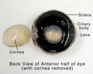 Source: learning-center.homesciencetools.com
The iris is the part of the eye that is colored. A cow s eye as normally provided will not be a neatly trimmed eyeball but a messy agglomeration of flesh. A tough clear covering over the iris and pupil that protects t. The front is responsible for the front part of the face of the cow. About press copyright contact us creators advertise developers terms privacy policy safety how youtube works test new features press copyright contact us creators.
 Source: chegg.com
Rods respond in dim light. The bundle of nerve fibers that carry information from the retina to the brain. If you continue browsing the site you agree to the use of cookies on this website. A thick jelly that helps give the eyeball its shape. Each eye has a blind spot where there are no light sensitive cells.
 Source: quizlet.com
A thick jelly that helps give the eyeball its shape. A thick jelly that helps give the eyeball its shape. These include eye sockets nasal cavity and mouth. Each eye has a blind spot where there are no light sensitive cells. The tapetum can be used for cows to see in the dark because the light can reflect on the tapetum so they can see in the dark.
 Source: pinterest.com
When a calf is born both parts of the skull are almost the same size. The bundle of nerve fibers that carry information from the retina to the brain. The tapetum can be used for cows to see in the dark because the light can reflect on the tapetum so they can see in the dark. The iris is the part of the eye that is colored. Rods respond in dim light.
 Source: pinterest.com
Thus the first job of the dissector is to remove the connective tissue to produce a neatly trimmed eyeball leaving the eyeball itself and the rear cord intact. Anatomy of a cow eye slideshare uses cookies to improve functionality and performance and to provide you with relevant advertising. Each eye has a blind spot where there are no light sensitive cells. Thus the first job of the dissector is to remove the connective tissue to produce a neatly trimmed eyeball leaving the eyeball itself and the rear cord intact. About press copyright contact us creators advertise developers terms privacy policy safety how youtube works test new features press copyright contact us creators.
If you find this site serviceableness, please support us by sharing this posts to your own social media accounts like Facebook, Instagram and so on or you can also bookmark this blog page with the title anatomy of a cow eye by using Ctrl + D for devices a laptop with a Windows operating system or Command + D for laptops with an Apple operating system. If you use a smartphone, you can also use the drawer menu of the browser you are using. Whether it’s a Windows, Mac, iOS or Android operating system, you will still be able to bookmark this website.

