Choroid layer of eye
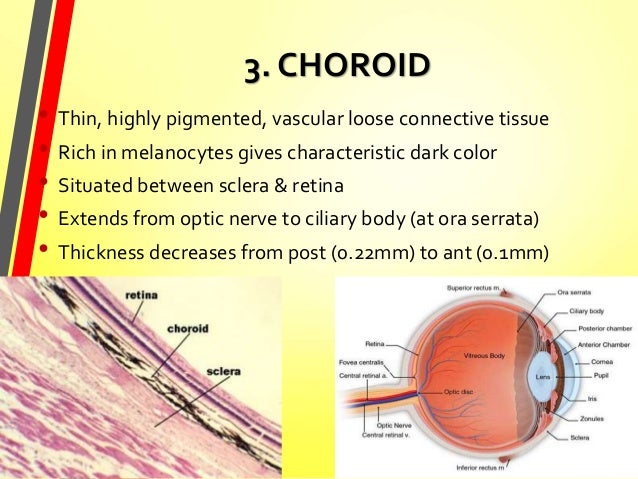
Choroid Layer Of Eye. The choroid is thickest in the back of the eye where it is about 0 2 mm and narrows to 0 1 mm in the peripheral part of the eye. The choroid layer also acts as a black screen which prevents extra reflections inside the eyeball so that we can get a perfect image. It is the vascular layer of the eye containing connective tissue and it nourishes the retina and absorbs scattered light. Its function is to provide nourishment to the outer layers of the retina through blood vessels.
 Anatomy Of The Eye Eye Physicians Of Lakewood David R Benson Md Od Pllc From eyecare-for-you.com
It joins the ciliary body anteriorly and lies between the retina and sclera posteriorly. The human choroid is thickest at the far extreme rear of the eye at 0 2 mm while in the outlying areas it narrows to 0 1 mm. Asked jul 3 in biology microbiology by galloway86. It consists of a densely capillary rich layer supplying blood to the eyeball. The choroid layer also acts as a black screen which prevents extra reflections inside the eyeball so that we can get a perfect image. 1 the choroid provides oxygen and nourishment to the outer layers of the retina.
Has an extensive blood supply.
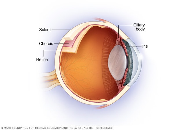
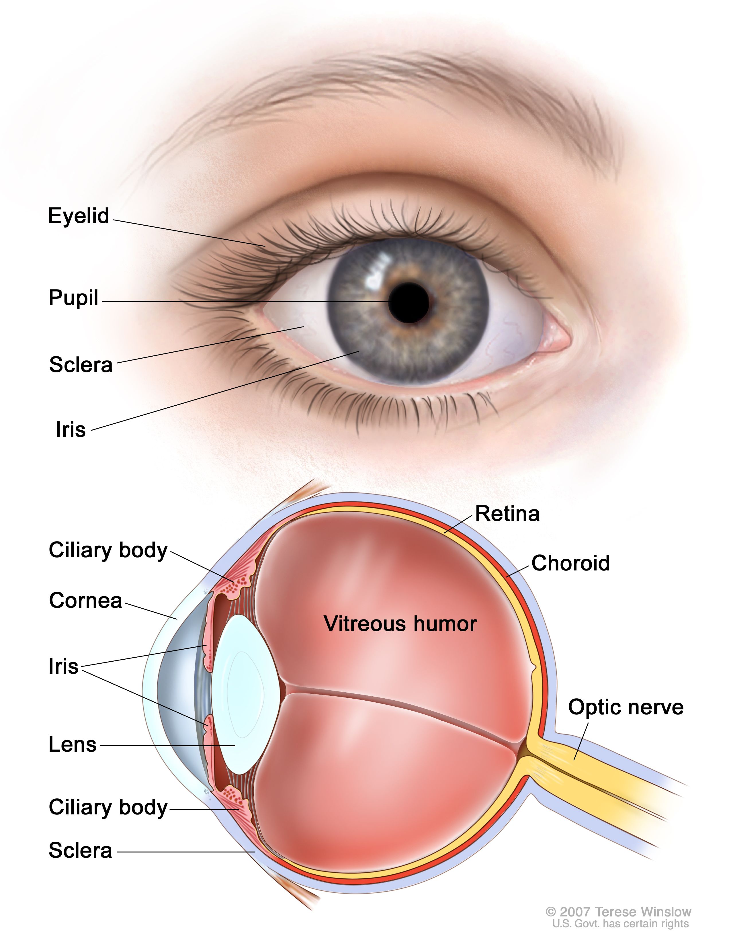
Has an extensive blood supply. It joins the ciliary body anteriorly and lies between the retina and sclera posteriorly. Contains a dark pigment. It is the vascular layer of the eye containing connective tissue and it nourishes the retina and absorbs scattered light. All of the above. The choroid is the middle layer of the eye that contains blood vessels and connective tissue between the sclera the white of the eye and the retina at the back of the eye.
 Source: eyecare-for-you.com
The choroid is a thin variably pigmented vascular tissue forming the posterior uvea. Asked jul 3 in biology microbiology by galloway86. My dashboard my education find an ophthalmologist. Has an extensive blood supply. The outer layer will form the substantia propria or stroma of the cornea.
Source: aao.org
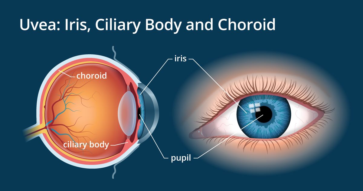
The inner layer is continuous with the choroid and is called the iridopupillary membrane and the outer layer is continuous with the sclera. The choroid layer also acts as a black screen which prevents extra reflections inside the eyeball so that we can get a perfect image. It contains the retinal pigmented epithelial cells and provides oxygen and nourishment to the outer retina. The choroid is part of the uvea and it contains blood vessels and connective tissue. My dashboard my education find an ophthalmologist.
 Source: allaboutvision.com
It joins the ciliary body anteriorly and lies between the retina and sclera posteriorly. Asked jul 3 in biology microbiology by galloway86. The outer layer will form the substantia propria or stroma of the cornea. Choroid is part of the uvea and supplies nutrients to the inner parts of the eye. The inner layer is continuous with the choroid and is called the iridopupillary membrane and the outer layer is continuous with the sclera.
 Source: en.wikipedia.org
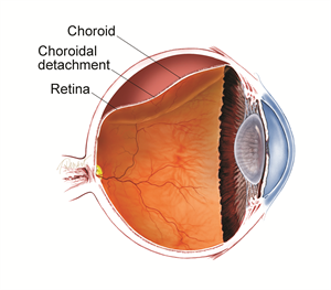
The choroid is the middle layer of the eye that contains blood vessels and connective tissue between the sclera the white of the eye and the retina at the back of the eye. The choroid also known as the choroidea or choroid coat is the vascular layer of the eye containing connective tissues and lying between the retina and the sclera. Its function is to provide nourishment to the outer layers of the retina through blood vessels. The vascular major blood vessel central layer of the eye lying between the retina and sclera. It is the vascular layer of the eye containing connective tissue and it nourishes the retina and absorbs scattered light.
 Source: asrs.org
My dashboard my education find an ophthalmologist. The choroid is the middle layer of the eye that contains blood vessels and connective tissue between the sclera the white of the eye and the retina at the back of the eye. The vascular major blood vessel central layer of the eye lying between the retina and sclera. The choroid is the vascular layer of the eye that lies between the retina and the sclera. Has an extensive blood supply.
 Source: en.wikipedia.org
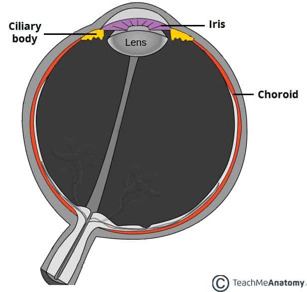
The choroid also known as the choroidea or choroid coat is the vascular layer of the eye containing connective tissues and lying between the retina and the sclera. This is the second layer forming the eyeball. All of the above. 1 the choroid provides oxygen and nourishment to the outer layers of the retina. Parts are the iris ciliary body and the ligaments.
 Source: pinterest.com
The vascular major blood vessel central layer of the eye lying between the retina and sclera. Parts are the iris ciliary body and the ligaments. Has an extensive blood supply. The outer layer will form the substantia propria or stroma of the cornea. It contains the retinal pigmented epithelial cells and provides oxygen and nourishment to the outer retina.
 Source: discovery.lifemapsc.com
The choroid is part of the uvea and it contains blood vessels and connective tissue. The cornea has three layers epithelium stroma and endothelium. The choroid is the vascular layer of the eye that lies between the retina and the sclera. The human choroid is thickest at the far extreme rear of the eye at 0 2 mm while in the outlying areas it narrows to 0 1 mm. This is the second layer forming the eyeball.
 Source: en.wikipedia.org
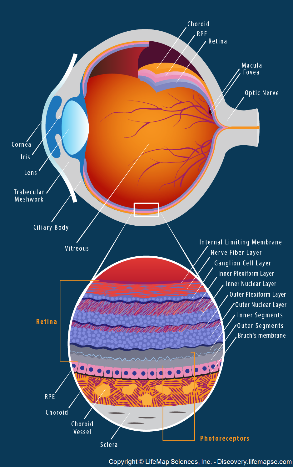
The choroid is extremely vascular with its capillaries arranged in a single layer on the inner surface to nourish the outer retinal layers figure 11 14. The human choroid is thickest at the far extreme rear of the eye at 0 2 mm while in the outlying areas it narrows to 0 1 mm. The cornea has three layers epithelium stroma and endothelium. This is the second layer forming the eyeball. The choroid is extremely vascular with its capillaries arranged in a single layer on the inner surface to nourish the outer retinal layers figure 11 14.
 Source: teachmeanatomy.info
The choroid is the middle layer between sclera and retina. The part of your eye between the sclera and the retina. The choroid is thickest in the back of the eye where it is about 0 2 mm and narrows to 0 1 mm in the peripheral part of the eye. The human choroid is thickest at the far extreme rear of the eye at 0 2 mm while in the outlying areas it narrows to 0 1 mm. The choroid is extremely vascular with its capillaries arranged in a single layer on the inner surface to nourish the outer retinal layers figure 11 14.
Source: researchgate.net
The choroid is the middle layer of the eye that contains blood vessels and connective tissue between the sclera the white of the eye and the retina at the back of the eye. The cornea has three layers epithelium stroma and endothelium. The choroid also known as the choroidea or choroid coat is the vascular layer of the eye containing connective tissues and lying between the retina and the sclera. Select the correct descriptions of the choroid layer of the human eye. The human choroid is thickest at the far extreme rear of the eye at 0 2 mm while in the outlying areas it narrows to 0 1 mm.
 Source: mayoclinic.org
This is the second layer forming the eyeball. Contains a dark pigment. Parts are the iris ciliary body and the ligaments. The choroid also known as the choroidea or choroid coat is the vascular layer of the eye containing connective tissues and lying between the retina and the sclera. The choroid layer also acts as a black screen which prevents extra reflections inside the eyeball so that we can get a perfect image.
 Source: slideshare.net
The part of your eye between the sclera and the retina. The choroid is the vascular layer of the eye that lies between the retina and the sclera. Asked jul 3 in biology microbiology by galloway86. It is the vascular layer of the eye containing connective tissue and it nourishes the retina and absorbs scattered light. The cornea has three layers epithelium stroma and endothelium.
 Source: researchgate.net
The choroid is extremely vascular with its capillaries arranged in a single layer on the inner surface to nourish the outer retinal layers figure 11 14. It contains the retinal pigmented epithelial cells and provides oxygen and nourishment to the outer retina. The outer layer will form the substantia propria or stroma of the cornea. The vascular major blood vessel central layer of the eye lying between the retina and sclera. The choroid is thickest in the back of the eye where it is about 0 2 mm and narrows to 0 1 mm in the peripheral part of the eye.
 Source: cancer.gov
The human choroid is thickest at the far extreme rear of the eye at 0 2 mm while in the outlying areas it narrows to 0 1 mm. The choroid is extremely vascular with its capillaries arranged in a single layer on the inner surface to nourish the outer retinal layers figure 11 14. The choroid also known as the choroidea or choroid coat is the vascular layer of the eye containing connective tissues and lying between the retina and the sclera. The outer layer will form the substantia propria or stroma of the cornea. Contains a dark pigment.
If you find this site beneficial, please support us by sharing this posts to your favorite social media accounts like Facebook, Instagram and so on or you can also save this blog page with the title choroid layer of eye by using Ctrl + D for devices a laptop with a Windows operating system or Command + D for laptops with an Apple operating system. If you use a smartphone, you can also use the drawer menu of the browser you are using. Whether it’s a Windows, Mac, iOS or Android operating system, you will still be able to bookmark this website.


