Colored threads under microscope
Colored Threads Under Microscope. Obtain a slide of colored threads and view them under the scanning and low power. It has a yellow thread on the bottom which is crossed by a blue thread which in turn is crossed by a red thread. I just wanted to know a little more about the threads i was using and reading about on other blogs. We know that the water bottle is behind the dna molecule.
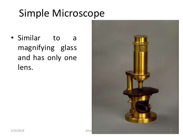
 Microscopy Htm From webpage.pace.edu
We know that the water bottle is behind the dna molecule. The left is. The improved clarity of the slide is worth it for teaching depth of field and focus. I decided the only way i could really tell the quality of a thread was to visually examine it feel it between my fingers and handle it in my machine. I just wanted to know a little more about the threads i was using and reading about on other blogs. This slide is used to help students learn to focus the microscope.
Each thread has depth and does not occupy the same exact space.
View the threads under high power not oil immersion. Each thread has depth and does not occupy the same exact space. Which is on bottom. I just wanted to know a little more about the threads i was using and reading about on other blogs. Which thread is on top. We recognize that they are on different planes because they are three dimensional.
 Source: us.vwr.com
Which is on bottom. Students use the fine focus to determine in which order the threads overlap. I just wanted to know a little more about the threads i was using and reading about on other blogs. It has a yellow thread on the bottom which is crossed by a blue thread which in turn is crossed by a red thread. Under the microscope the threads of differing color are also stacked on top of each other.
 Source: amazon.com
I just wanted to know a little more about the threads i was using and reading about on other blogs. Use the focusing procedure described above. Use the fine focus to figure out the order of the threads from top to bottom. Each thread has depth and does not occupy the same exact space. Which is on bottom.
 Source: bio100.nicerweb.com
Which is on bottom. Which thread is on top. In this article i will share with you what your thread looks like under a microscope. Prepared slide of colored threads under the microscope. It has a yellow thread on the bottom which is crossed by a blue thread which in turn is crossed by a red thread.
 Source: homesciencetools.com
The improved clarity of the slide is worth it for teaching depth of field and focus. It has a yellow thread on the bottom which is crossed by a blue thread which in turn is crossed by a red thread. I just wanted to know a little more about the threads i was using and reading about on other blogs. One of the key things i learned from this experiment was that the letter under the microscope looks not only bigger but is reflected as though the view is under the microscope and is upside down. I decided the only way i could really tell the quality of a thread was to visually examine it feel it between my fingers and handle it in my machine.
 Source: m.youtube.com
Obtain a slide of colored threads and view them under the scanning power. I just wanted to know a little more about the threads i was using and reading about on other blogs. View the threads under high power 400x or 430x. View the threads under high power not oil immersion. I decided the only way i could really tell the quality of a thread was to visually examine it feel it between my fingers and handle it in my machine.
 Source: pinterest.com
View the threads under high power not oil immersion. In this article i will share with you what your thread looks like under a microscope. I decided the only way i could really tell the quality of a thread was to visually examine it feel it between my fingers and handle it in my machine. Use the fine focus to figure out the order of the threads from top to bottom. View the threads under high power not oil immersion.
 Source: thehappyscientist.com
Use the focusing procedure described above. The improved clarity of the slide is worth it for teaching depth of field and focus. Use the fine focus to figure out the order of the threads from top to bottom. Obtain a slide of colored threads and view them under the scanning and low power. If you are working with partners on this exercise be sure that everybody in your group uses the microscope.
Source: webpage.pace.edu
This whole mount of 3 different colored threads is perfect when teaching your students how to focus the compound microscope. It has a yellow thread on the bottom which is crossed by a blue thread which in turn is crossed by a red thread. Which is on bottom. Use the fine focus to figure out the order of the threads from top to bottom. Each thread has depth and does not occupy the same exact space.
 Source: learningsurge.com
Students use the fine focus to determine in which order the threads overlap. This slide is used to help students learn to focus the microscope. It has a yellow thread on the bottom which is crossed by a blue thread which in turn is crossed by a red thread. In this article i will share with you what your thread looks like under a microscope. We know that the water bottle is behind the dna molecule.
 Source: williambiolabreports.wordpress.com
One of the key things i learned from this experiment was that the letter under the microscope looks not only bigger but is reflected as though the view is under the microscope and is upside down. Which is on bottom. We know that the water bottle is behind the dna molecule. This whole mount of 3 different colored threads is perfect when teaching your students how to focus the compound microscope. Use the fine focus to figure out the order of the threads from top to bottom.
 Source: sites.google.com
1 can you tell which thread is above the other. Use the fine focus to figure out the order of the threads from top to bottom. Students use the fine focus to determine in which order the threads overlap. Under the microscope the threads of differing color are also stacked on top of each other. Obtain a slide of colored threads and view them under the scanning and low power.
 Source: sciences.usca.edu
Prepared slide of colored threads under the microscope. We recognize that they are on different planes because they are three dimensional. We know that the water bottle is behind the dna molecule. Obtain a slide of colored threads and view them under the scanning and low power. Students use the fine focus to determine in which order the threads overlap.
 Source: slideplayer.com
We recognize that they are on different planes because they are three dimensional. Obtain a slide of colored threads and view them under the scanning and low power. 1 can you tell which thread is above the other. In this article i will share with you what your thread looks like under a microscope. Each thread has depth and does not occupy the same exact space.
 Source: taylor-biology100.weebly.com
View the threads under high power 400x or 430x. 1 can you tell which thread is above the other. The left is. Which thread is on top. I just wanted to know a little more about the threads i was using and reading about on other blogs.
 Source: steemit.com
Use the focusing procedure described above. Prepared slide of colored threads under the microscope. Use the fine focus to figure out the order of the threads from top to bottom. This slide is used to help students learn to focus the microscope. The improved clarity of the slide is worth it for teaching depth of field and focus.
If you find this site adventageous, please support us by sharing this posts to your own social media accounts like Facebook, Instagram and so on or you can also save this blog page with the title colored threads under microscope by using Ctrl + D for devices a laptop with a Windows operating system or Command + D for laptops with an Apple operating system. If you use a smartphone, you can also use the drawer menu of the browser you are using. Whether it’s a Windows, Mac, iOS or Android operating system, you will still be able to bookmark this website.