Cow eye anatomy
Cow Eye Anatomy. Obtain a cow eye dissection styrofoam tray gloves and surgical tools. The colorful shiny material located behind the retina. A cow s eye as normally provided will not be a neatly trimmed eyeball but a messy agglomeration of flesh. Leave the optic nerve attached.
 Cow Eye Dissection Cow Eyes Eye Structure Eyeball Diagram From pinterest.com
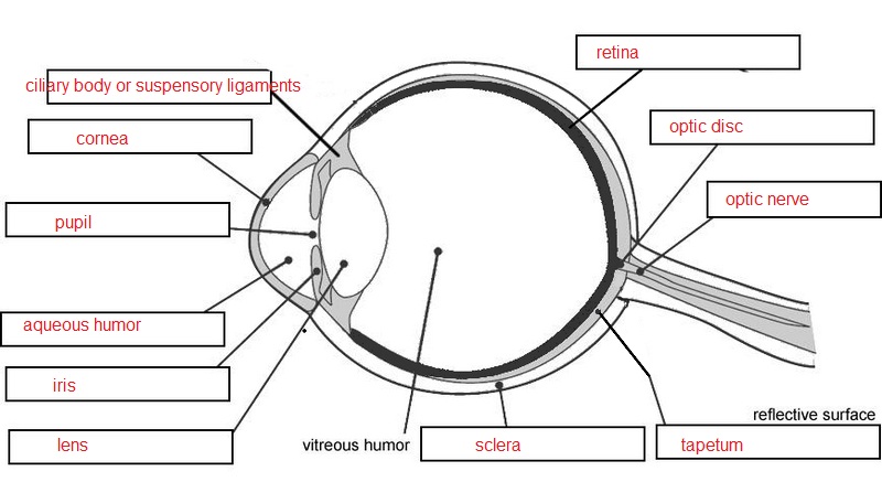
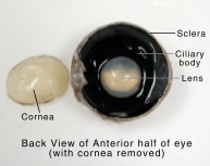
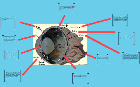
A dark hole in the center of the iris that lets light into the. The most noticeable part of the eye is the large mass of gray tissue that surrounds the posterior back of the eye and is attached to the sclera. Cut the eyeball in half. The bundle of nerve fibers that carry information from the retina to the brain. A cow s eye as normally provided will not be a neatly trimmed eyeball but a messy agglomeration of flesh. Found in animals with good night vision the tapetum reflects light back through the retina.
About press copyright contact us creators advertise developers terms privacy policy safety how youtube works test new features press copyright contact us creators.
With a sharp point poke a whole on the side of the eye to relieve the pressure in the eyeball so you wont get squirted with liquid when cutting open the eyeball. A thick jelly that helps give the eyeball its shape. Remove excess tissue from the eye such as the fat muscle eye lids etc. Cut the eyeball in half. Obtain a cow eye dissection styrofoam tray gloves and surgical tools. Found in animals with good night vision the tapetum reflects light back through the retina.
 Source: pinterest.com
About press copyright contact us creators advertise developers terms privacy policy safety how youtube works test new features press copyright contact us creators. The second most noticeable part of the eye is the cornea located in the anterior front part of the eye. A thick jelly that helps give the eyeball its shape. With a sharp point poke a whole on the side of the eye to relieve the pressure in the eyeball so you wont get squirted with liquid when cutting open the eyeball. Leave the optic nerve attached.
 Source: quizlet.com
A dark hole in the center of the iris that lets light into the. A tough clear covering over the iris and pupil that protects t. The bundle of nerve fibers that carry information from the retina to the brain. A dark hole in the center of the iris that lets light into the. About press copyright contact us creators advertise developers terms privacy policy safety how youtube works test new features press copyright contact us creators.
 Source: quizlet.com
Obtain a cow eye dissection styrofoam tray gloves and surgical tools. The most noticeable part of the eye is the large mass of gray tissue that surrounds the posterior back of the eye and is attached to the sclera. With a sharp point poke a whole on the side of the eye to relieve the pressure in the eyeball so you wont get squirted with liquid when cutting open the eyeball. About press copyright contact us creators advertise developers terms privacy policy safety how youtube works test new features press copyright contact us creators. Take pictures of each step of the eye process.
 Source: biologyjunction.com
Found in animals with good night vision the tapetum reflects light back through the retina. Cut the eyeball in half. Thus the first job of the dissector is to remove the connective tissue to produce a neatly trimmed eyeball leaving the eyeball itself and the rear cord intact. With a sharp point poke a whole on the side of the eye to relieve the pressure in the eyeball so you wont get squirted with liquid when cutting open the eyeball. Found in animals with good night vision the tapetum reflects light back through the retina.
 Source: science.jburroughs.org
Remove excess tissue from the eye such as the fat muscle eye lids etc. About press copyright contact us creators advertise developers terms privacy policy safety how youtube works test new features press copyright contact us creators. The bundle of nerve fibers that carry information from the retina to the brain. About press copyright contact us creators advertise developers terms privacy policy safety how youtube works test new features press copyright contact us creators. The most noticeable part of the eye is the large mass of gray tissue that surrounds the posterior back of the eye and is attached to the sclera.
 Source: pinterest.com
The second most noticeable part of the eye is the cornea located in the anterior front part of the eye. The bundle of nerve fibers that carry information from the retina to the brain. The thick tough white outer covering of the eyeball. A thick jelly that helps give the eyeball its shape. Found in animals with good night vision the tapetum reflects light back through the retina.
 Source: biologycorner.com
Leave the optic nerve attached. Obtain a cow eye dissection styrofoam tray gloves and surgical tools. The bundle of nerve fibers that carry information from the retina to the brain. The most noticeable part of the eye is the large mass of gray tissue that surrounds the posterior back of the eye and is attached to the sclera. Found in animals with good night vision the tapetum reflects light back through the retina.
 Source: learning-center.homesciencetools.com
With a sharp point poke a whole on the side of the eye to relieve the pressure in the eyeball so you wont get squirted with liquid when cutting open the eyeball. Look carefully at the preserved cow eye. A tough clear covering over the iris and pupil that protects t. The optic nerve s job is to send information from that was accumulated in the eye to the brain so you can understand what you are looking at. Obtain a cow eye dissection styrofoam tray gloves and surgical tools.
 Source: homesciencetools.com
The colorful shiny material located behind the retina. A dark hole in the center of the iris that lets light into the. With a sharp point poke a whole on the side of the eye to relieve the pressure in the eyeball so you wont get squirted with liquid when cutting open the eyeball. Although they do not show it here there is one more part of the eye called the optic nerve that lies on the anterior of the eye. Remove excess tissue from the eye such as the fat muscle eye lids etc.
 Source: prezi.com
The optic nerve s job is to send information from that was accumulated in the eye to the brain so you can understand what you are looking at. Look carefully at the preserved cow eye. A thick jelly that helps give the eyeball its shape. Take pictures of each step of the eye process. Leave the optic nerve attached.
 Source: anatomycorner.com
The most noticeable part of the eye is the large mass of gray tissue that surrounds the posterior back of the eye and is attached to the sclera. The bundle of nerve fibers that carry information from the retina to the brain. Leave the optic nerve attached. With a sharp point poke a whole on the side of the eye to relieve the pressure in the eyeball so you wont get squirted with liquid when cutting open the eyeball. Remove excess tissue from the eye such as the fat muscle eye lids etc.
 Source: chegg.com
Remove excess tissue from the eye such as the fat muscle eye lids etc. Take pictures of each step of the eye process. The thick tough white outer covering of the eyeball. A thick jelly that helps give the eyeball its shape. Remove excess tissue from the eye such as the fat muscle eye lids etc.
 Source: pinterest.com
Found in animals with good night vision the tapetum reflects light back through the retina. A tough clear covering over the iris and pupil that protects t. The second most noticeable part of the eye is the cornea located in the anterior front part of the eye. Remove excess tissue from the eye such as the fat muscle eye lids etc. Leave the optic nerve attached.
 Source: pinterest.com
The second most noticeable part of the eye is the cornea located in the anterior front part of the eye. The most noticeable part of the eye is the large mass of gray tissue that surrounds the posterior back of the eye and is attached to the sclera. Cut the eyeball in half. The optic nerve s job is to send information from that was accumulated in the eye to the brain so you can understand what you are looking at. The bundle of nerve fibers that carry information from the retina to the brain.
 Source: pinterest.com
Look carefully at the preserved cow eye. Found in animals with good night vision the tapetum reflects light back through the retina. A cow s eye as normally provided will not be a neatly trimmed eyeball but a messy agglomeration of flesh. With a sharp point poke a whole on the side of the eye to relieve the pressure in the eyeball so you wont get squirted with liquid when cutting open the eyeball. Cut the eyeball in half.
If you find this site value, please support us by sharing this posts to your preference social media accounts like Facebook, Instagram and so on or you can also save this blog page with the title cow eye anatomy by using Ctrl + D for devices a laptop with a Windows operating system or Command + D for laptops with an Apple operating system. If you use a smartphone, you can also use the drawer menu of the browser you are using. Whether it’s a Windows, Mac, iOS or Android operating system, you will still be able to bookmark this website.


