Internal anatomy of a fetal pig labeled
Internal Anatomy Of A Fetal Pig Labeled. Phylum chordata subphylum vertebrat. The thoracic cavity and the abdominal cavity are separated by the diaphragm. This is a gray ish mass that covers the upper trachea. It is opposite the dorsal side.
 Reading Fetal Pig Dissection Biology Ii Laboratory Manual From courses.lumenlearning.com
The pig in the first photograph below has its ventral side up. This is a gray ish mass that covers the upper trachea. Ventral is the belly side. The thoracic cavity and the abdominal cavity are separated by the diaphragm. The thoracic cavity which contains the lungs and the abdominal cavity which contains the digestive system. Internal anatomy of a fetal pig emphasizing the cardiorespiratory digestive and urogenital systems male and female.
The pig in the first photograph below has its ventral side up.
The thoracic cavity which contains the lungs and the abdominal cavity which contains the digestive system. Internal anatomy of a fetal pig emphasizing the cardiorespiratory digestive and urogenital systems male and female. It is opposite the dorsal side. Using the diagram to the left. Phylum chordata subphylum vertebrat. Use the photographs below to identify its sex.
 Source:
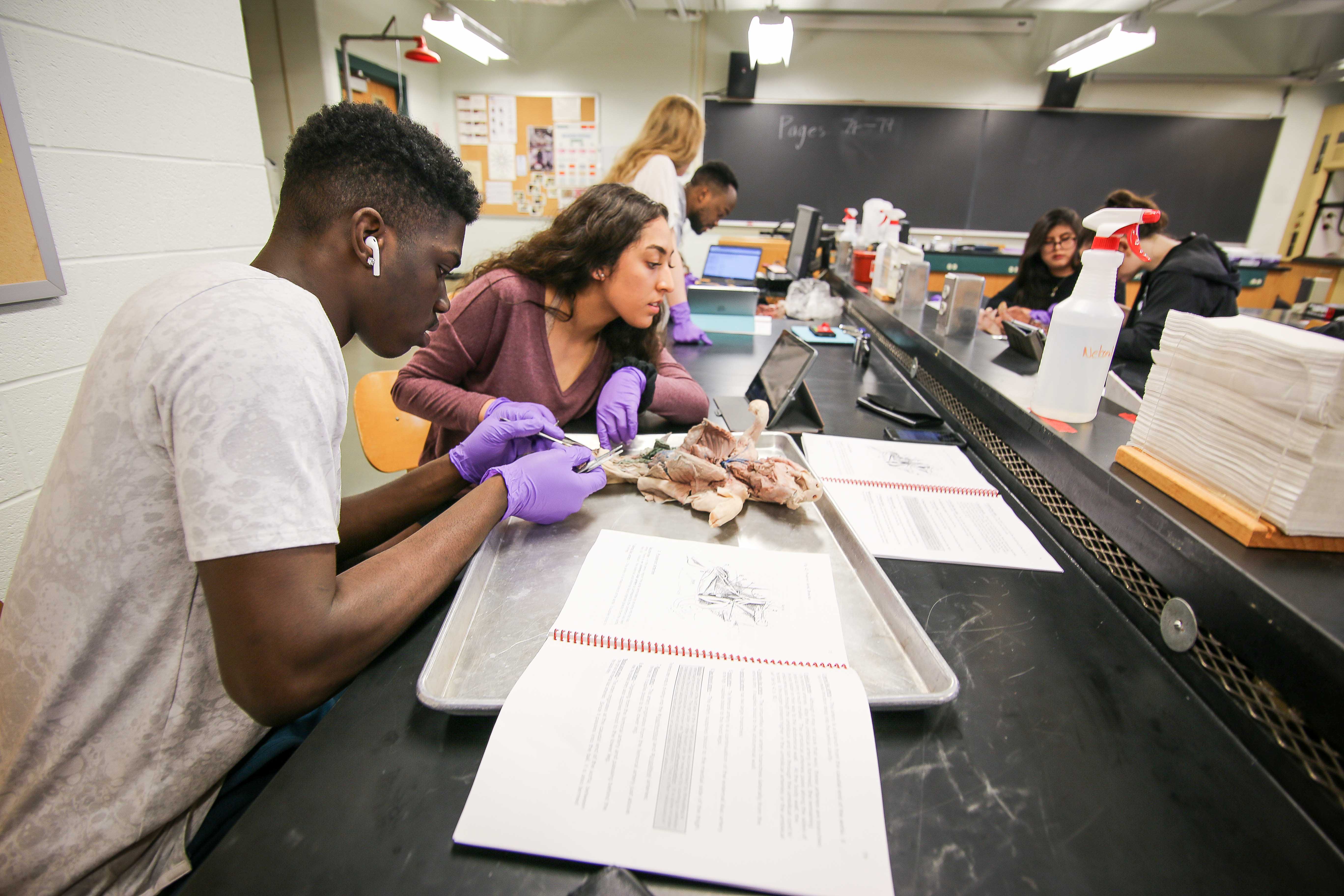
Obtain a fetal pig and identify the structures listed in the first photograph. Identify two bean shaped kidneys the dorsal wall in the mid to lower back region the light coloured strip of tissue at the top of each kidney is an adrenal gland. The thoracic cavity and the abdominal cavity are separated by the diaphragm. The pig in the first photograph below is laying on its dorsal side. Larynx the structure of cartilage contains the vocal chords.
 Source: pinterest.com
The pig in the first photograph below has its ventral side up. This is a gray ish mass that covers the upper trachea. It is opposite the dorsal side. In mammals the coelom is divided into two main cavities. The pig in the first photograph below is laying on its dorsal side.
Source: courses.lumenlearning.com
This is a gray ish mass that covers the upper trachea. Note the many membranes lining the coelom and holding the organs in place. In mammals the coelom is divided into two main cavities. Ventral is the belly side. Phylum chordata subphylum vertebrat.
 Source: sdmesa.edu
Larynx the structure of cartilage contains the vocal chords. The thoracic cavity which contains the lungs and the abdominal cavity which contains the digestive system. Use the photographs below to identify its sex. The pig in the first photograph below is laying on its dorsal side. Note the many membranes lining the coelom and holding the organs in place.
 Source: biologycorner.com
The thoracic cavity which contains the lungs and the abdominal cavity which contains the digestive system. In mammals the coelom is divided into two main cavities. Internal anatomy of a fetal pig. Internal anatomy of a fetal pig emphasizing the cardiorespiratory digestive and urogenital systems male and female. Identify two bean shaped kidneys the dorsal wall in the mid to lower back region the light coloured strip of tissue at the top of each kidney is an adrenal gland.
 Source: frogdiagrams.ammediocasa.it
Phylum chordata subphylum vertebrat. Larynx the structure of cartilage contains the vocal chords. Internal anatomy of a fetal pig emphasizing the cardiorespiratory digestive and urogenital systems male and female. Use the photographs below to identify its sex. The pig in the first photograph below has its ventral side up.
 Source: pinterest.com
The pig in the first photograph below has its ventral side up. Identify the ureters which carry urine from the kidneys to the urinary bladder find the urinary bladder within the umbillical cord. The thoracic cavity and the abdominal cavity are separated by the diaphragm. Identify two bean shaped kidneys the dorsal wall in the mid to lower back region the light coloured strip of tissue at the top of each kidney is an adrenal gland. Phylum chordata subphylum vertebrat.
Source:
Identify the ureters which carry urine from the kidneys to the urinary bladder find the urinary bladder within the umbillical cord. In mammals the coelom is divided into two main cavities. Obtain a fetal pig and identify the structures listed in the first photograph. This is a gray ish mass that covers the upper trachea. Identify two bean shaped kidneys the dorsal wall in the mid to lower back region the light coloured strip of tissue at the top of each kidney is an adrenal gland.
 Source: biologyjunction.com
Note the many membranes lining the coelom and holding the organs in place. Use the photographs below to identify its sex. Ventral is the belly side. Phylum chordata subphylum vertebrat. Larynx the structure of cartilage contains the vocal chords.
 Source: br.pinterest.com
The thoracic cavity and the abdominal cavity are separated by the diaphragm. Identify the ureters which carry urine from the kidneys to the urinary bladder find the urinary bladder within the umbillical cord. It is opposite the dorsal side. This is a gray ish mass that covers the upper trachea. Obtain a fetal pig and identify the structures listed in the first photograph.
 Source: biologycorner.com
The pig in the first photograph below has its ventral side up. Identify the ureters which carry urine from the kidneys to the urinary bladder find the urinary bladder within the umbillical cord. Use the photographs below to identify its sex. Larynx the structure of cartilage contains the vocal chords. Internal anatomy of a fetal pig emphasizing the cardiorespiratory digestive and urogenital systems male and female.
 Source: m.youtube.com
Note the many membranes lining the coelom and holding the organs in place. Phylum chordata subphylum vertebrat. The thoracic cavity and the abdominal cavity are separated by the diaphragm. Using the diagram to the left. Identify the ureters which carry urine from the kidneys to the urinary bladder find the urinary bladder within the umbillical cord.
 Source: courses.lumenlearning.com
Use the photographs below to identify its sex. Phylum chordata subphylum vertebrat. Internal anatomy of a fetal pig. Internal anatomy of a fetal pig emphasizing the cardiorespiratory digestive and urogenital systems male and female. In mammals the coelom is divided into two main cavities.
Source:
The pig in the first photograph below has its ventral side up. This is a gray ish mass that covers the upper trachea. The pig in the first photograph below has its ventral side up. This is a gray ish mass that covers the upper trachea. The pig in the first photograph below is laying on its dorsal side.
 Source: biologycorner.com
This is a gray ish mass that covers the upper trachea. Obtain a fetal pig and identify the structures listed in the first photograph. This is a gray ish mass that covers the upper trachea. Internal anatomy of a fetal pig. Identify two bean shaped kidneys the dorsal wall in the mid to lower back region the light coloured strip of tissue at the top of each kidney is an adrenal gland.
If you find this site value, please support us by sharing this posts to your favorite social media accounts like Facebook, Instagram and so on or you can also bookmark this blog page with the title internal anatomy of a fetal pig labeled by using Ctrl + D for devices a laptop with a Windows operating system or Command + D for laptops with an Apple operating system. If you use a smartphone, you can also use the drawer menu of the browser you are using. Whether it’s a Windows, Mac, iOS or Android operating system, you will still be able to bookmark this website.