Layers of the eyeball
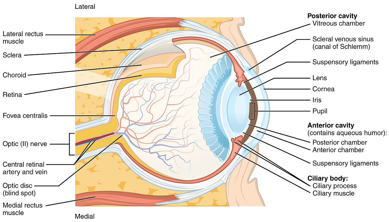
Layers Of The Eyeball. It consists of the sclera and cornea which are continuous with each other. The vascular layer protects portions of the eye. At the front of the eyeball the sclera becomes the cornea. The iris choroid and ciliary body.
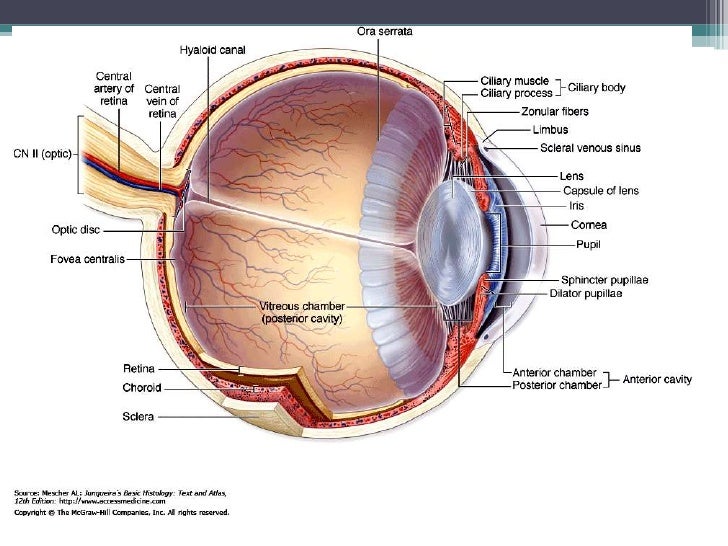 Eye Layers 1 From slideshare.net
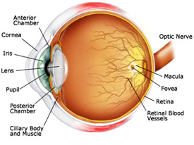
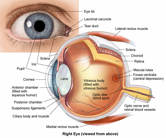
The human eye is a marvel of anatomy providing us with the ability to see the world in all its textures colors and sizes. Diagram of the different layers of the eyeball. Their main functions are to provide shape to the eye and support the deeper structures. The lacrimal gland produces tears that help lubricate and moisten the eye as well as flush away any foreign matter that may enter the eye. The human eye has three layers of eye tissue. The iris choroid and ciliary body.
It consists of the sclera and cornea which are continuous with each other.
It is the white and opaque part of the eyeball. The cornea is the transparent dome shaped part of the eyeball. While dividing the eye into multiple layers can be done in a variety of ways one way to think of layers of the eye is to consider the eyeball as being composed of three main layers. It consists of the sclera and cornea which are continuous with each other. The spaces within the eye are filled with fluids that help maintain its shape. The sclera is outermost layer of the eyeball.
 Source: youtube.com
Tears lubricate the eye and are made up of three layers. Tears lubricate the eye and are made up of three layers. The orbit also contains the lacrimal gland that is located underneath the outer portion of the upper eyelid. The fibrous layer of the eye is the outermost layer. It is the white and opaque part of the eyeball.
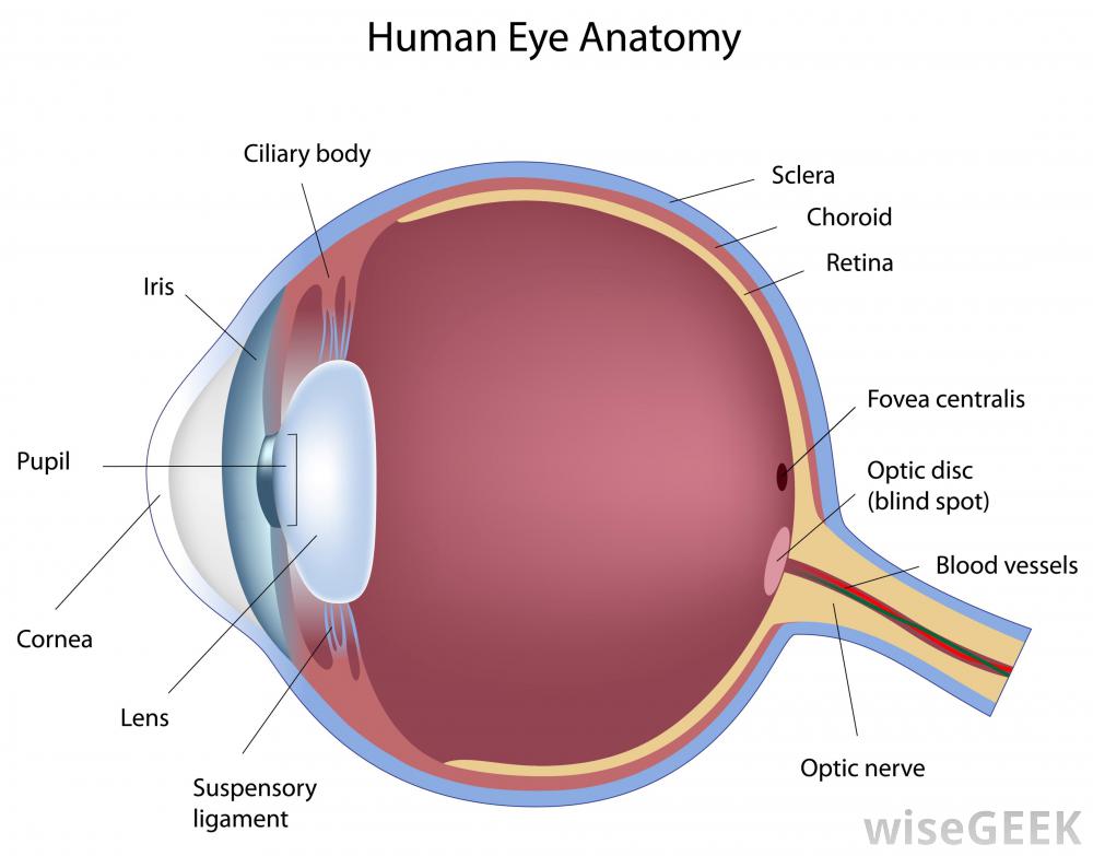 Source: socratic.org
The iris choroid and ciliary body. While dividing the eye into multiple layers can be done in a variety of ways one way to think of layers of the eye is to consider the eyeball as being composed of three main layers. This is a strong layer of tissue that covers nearly the entire surface of the eyeball. Wall of the eyeball the wall of the eyeball is made up of three layers fibrous outer vascular muscular middle and sensorineural inner layers. The iris choroid and ciliary body.
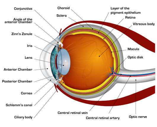 Source: nkcf.org
It consists of the sclera and cornea which are continuous with each other. Each of these layers performs a different function in helping a human see or in eye as well as forms a place for muscles to attach to. The eyeball can be divided into the fibrous vascular and inner layers. It is the white and opaque part of the eyeball. While dividing the eye into multiple layers can be done in a variety of ways one way to think of layers of the eye is to consider the eyeball as being composed of three main layers.
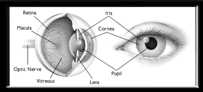 Source: ottawahospital.on.ca
Wall of the eyeball the wall of the eyeball is made up of three layers fibrous outer vascular muscular middle and sensorineural inner layers. Each of these layers performs a different function in helping a human see or in eye as well as forms a place for muscles to attach to. The fibrous layer the vascular layer and the retina. The eye is a hollow spherical structure about 2 5 centimeters in diameter. Muscles responsible for moving the eyeball are attached to the eyeball at the sclera.
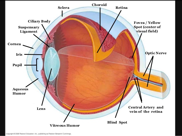 Source: socratic.org
These three layers together are called the tear film. The cornea is the transparent dome shaped part of the eyeball. Muscles responsible for moving the eyeball are attached to the eyeball at the sclera. The lacrimal gland produces tears that help lubricate and moisten the eye as well as flush away any foreign matter that may enter the eye. Fibrous vascular pigmented and nervous retina.
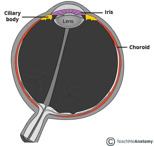 Source: teachmeanatomy.info
Wall of the eyeball the wall of the eyeball is made up of three layers fibrous outer vascular muscular middle and sensorineural inner layers. It consists of the sclera and cornea which are continuous with each other. Fibrous vascular pigmented and nervous retina. The eyeball can be divided into the fibrous vascular and inner layers. The spaces within the eye are filled with fluids that help maintain its shape.
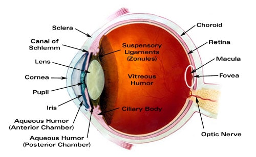 Source: laramyk.com
It consists of the sclera and cornea which are continuous with each other. It consists of the sclera and cornea which are continuous with each other. Functionally the most important layer is the retina which receives the external visual stimuli. It is the white and opaque part of the eyeball. The cornea and sclera.
 Source: slideshare.net
This is a strong layer of tissue that covers nearly the entire surface of the eyeball. Muscles responsible for moving the eyeball are attached to the eyeball at the sclera. While dividing the eye into multiple layers can be done in a variety of ways one way to think of layers of the eye is to consider the eyeball as being composed of three main layers. These three layers together are called the tear film. Wall of the eyeball the wall of the eyeball is made up of three layers fibrous outer vascular muscular middle and sensorineural inner layers.
 Source: in.pinterest.com
The vascular layer protects portions of the eye. The sclera is outermost layer of the eyeball. The eye is a hollow spherical structure about 2 5 centimeters in diameter. Fibrous vascular pigmented and nervous retina. Muscles responsible for moving the eyeball are attached to the eyeball at the sclera.
 Source: content.byui.edu
The surface of the eye and the inner surface of the eyelids are covered with a clear membrane called the conjunctiva. The cornea and sclera. The fibrous layer the vascular layer and the retina. The orbit also contains the lacrimal gland that is located underneath the outer portion of the upper eyelid. It is the white and opaque part of the eyeball.
 Source: birlaeye.org
The fibrous layer of the eye is the outermost layer. Each of these layers performs a different function in helping a human see or in eye as well as forms a place for muscles to attach to. The orbit also contains the lacrimal gland that is located underneath the outer portion of the upper eyelid. The vascular layer protects portions of the eye. We shall now look at these layers in further detail.
 Source: commons.wikimedia.org
Its wall has three distinct layers an outer fibrous layer a middle vascular layer and an inner nervous layer. The eyeball consists of three layers. It consists of the sclera and cornea which are continuous with each other. The cornea and sclera. The human eye is a marvel of anatomy providing us with the ability to see the world in all its textures colors and sizes.
 Source: youreyescenter.com
The orbit also contains the lacrimal gland that is located underneath the outer portion of the upper eyelid. Each of these layers performs a different function in helping a human see or in eye as well as forms a place for muscles to attach to. Anatomy of the eye. Diagram of the different layers of the eyeball. The human eye is a marvel of anatomy providing us with the ability to see the world in all its textures colors and sizes.
 Source: ophthalmologyforhealthcareassistants.wordpress.com
Muscles responsible for moving the eyeball are attached to the eyeball at the sclera. Their main functions are to provide shape to the eye and support the deeper structures. The cornea is the transparent dome shaped part of the eyeball. These layers have different structures and functions. It is the white and opaque part of the eyeball.
Source: researchgate.net
Muscles responsible for moving the eyeball are attached to the eyeball at the sclera. These layers have different structures and functions. The lacrimal gland produces tears that help lubricate and moisten the eye as well as flush away any foreign matter that may enter the eye. The fibrous layer the vascular layer and the retina. Each of these layers performs a different function in helping a human see or in eye as well as forms a place for muscles to attach to.
If you find this site beneficial, please support us by sharing this posts to your preference social media accounts like Facebook, Instagram and so on or you can also save this blog page with the title layers of the eyeball by using Ctrl + D for devices a laptop with a Windows operating system or Command + D for laptops with an Apple operating system. If you use a smartphone, you can also use the drawer menu of the browser you are using. Whether it’s a Windows, Mac, iOS or Android operating system, you will still be able to bookmark this website.


