Microscope iris diaphragm
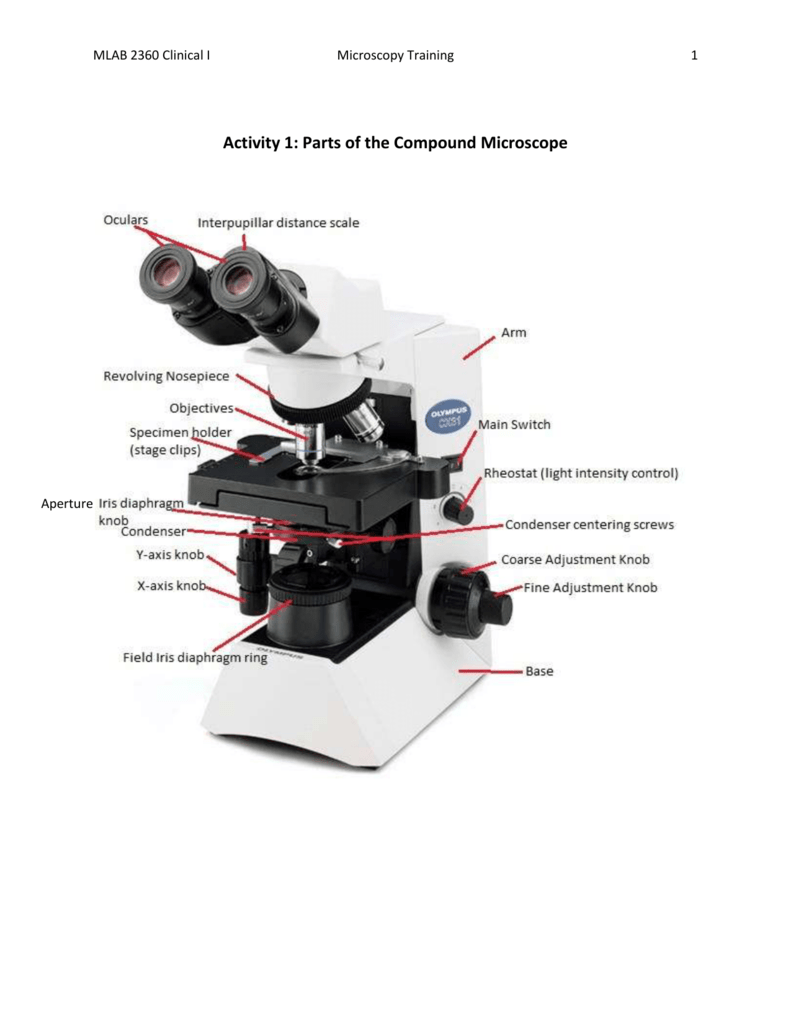
Microscope Iris Diaphragm. Most high quality microscopes include an abbe condenser with an iris diaphragm. It s aim is to improve beam shape in condenser system or image contrast in objective system the diaphragm or iris location is under the stage of the microscope and above it is the condenser this apparatus function is to control the amount of light that is allowed to pass through the specimen being viewed most quality microscopes. It operates in the same fashion as described above but it serves a little different purpose then just making the image more or less bright. In a microscope an iris diaphragm is an important component that directly influences the amount of illumination focus and contrast of the magnified specimen image.
 Microscope World Blog Microscope Disc Diaphragm From blog.microscopeworld.com
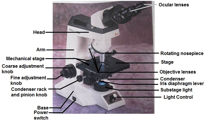
In fact the condenser sits right on top of the iris diaphragm. This is done by a rotating disc under the stage that has different sized holes for the light to shine through. Equipped on the condenser of the microscope the iris diaphragm is a shutter controlled by a lever that is used to regulate the amount of light entering the lens system. The general rule is the diaphragm s aperture size is directly proportional to illumination and conversely proportional to contrast while the aperture shape is directly proportional to focus. Most high quality microscopes include an abbe condenser with an iris diaphragm. In a microscope an iris diaphragm is an important component that directly influences the amount of illumination focus and contrast of the magnified specimen image.
Click to see full answer.
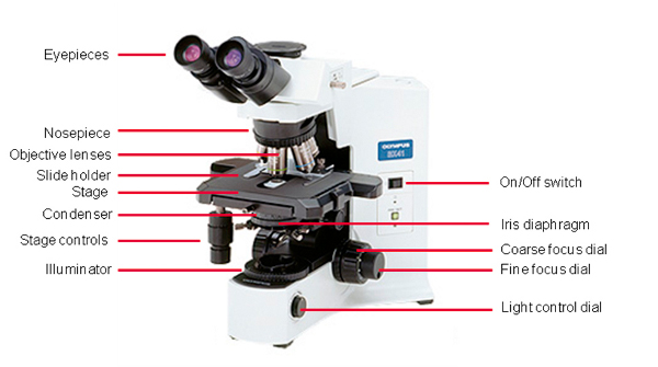
Combined they control both the focus and quantity of light applied to the specimen. The field iris diaphragm residing in a conjugate plane with the lamp collector lens is imaged sharply into the same plane as the specimen by the microscope condenser. Most high quality microscopes include an abbe condenser with an iris diaphragm. The diaphragm is located directly under the stage or platform where user places the specimen or slide. The diaphragm also called iris is located under the stage and is used to adjust and change the intensity and size of the cone of light that shines up through the side. Images of both the field diaphragm and the specimen are formed in the intermediate image plane by the objective and are projected into the fixed field diaphragm of the eyepiece where the focusing reticle is located.
 Source: amazon.com
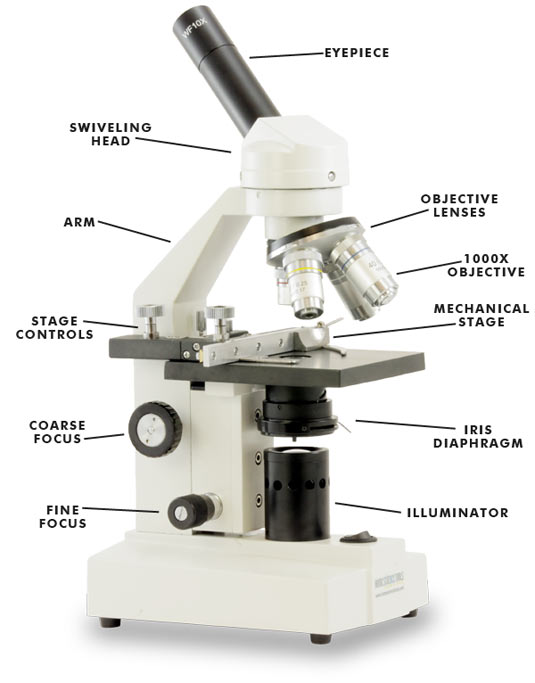
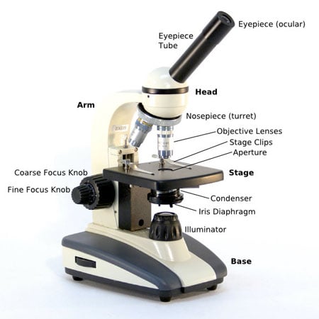
In a microscope an iris diaphragm is an important component that directly influences the amount of illumination focus and contrast of the magnified specimen image. Combined they control both the focus and quantity of light applied to the specimen. Closing the iris diaphragm will reduce the amount of illumination of the specimen but increases the amount of contrast. It is located above the condenser and below the stage. The diaphragm also called iris is located under the stage and is used to adjust and change the intensity and size of the cone of light that shines up through the side.
 Source: precisemicroscope.com
As in how much light and dark differ from each other in an image. By opening the diaphragm an item that at first appears too dark is easier to observe. In light microscopy the iris diaphragm controls the size of the opening between the specimen and condenser through which light passes. As in how much light and dark differ from each other in an image. This diaphragm is located closer to the condenser system of a microscope.
 Source: blog.microscopeworld.com
Images of both the field diaphragm and the specimen are formed in the intermediate image plane by the objective and are projected into the fixed field diaphragm of the eyepiece where the focusing reticle is located. This diaphragm is mostly used to control the contrast. Combined they control both the focus and quantity of light applied to the specimen. Iris diaphragm controls the amount of light reaching the specimen. Click to see full answer.
 Source: bitesizebio.com
It s aim is to improve beam shape in condenser system or image contrast in objective system the diaphragm or iris location is under the stage of the microscope and above it is the condenser this apparatus function is to control the amount of light that is allowed to pass through the specimen being viewed most quality microscopes. Iris diaphragm controls the amount of light reaching the specimen. In light microscopy the iris diaphragm controls the size of the opening between the specimen and condenser through which light passes. In fact the condenser sits right on top of the iris diaphragm. Combined they control both the focus and quantity of light applied to the specimen.
 Source: biologyaspoetry.com
In light microscopy the iris diaphragm controls the size of the opening between the specimen and condenser through which light passes. Closing the iris diaphragm will reduce the amount of illumination of the specimen but increases the amount of contrast. Equipped on the condenser of the microscope the iris diaphragm is a shutter controlled by a lever that is used to regulate the amount of light entering the lens system. By opening the diaphragm an item that at first appears too dark is easier to observe. This is done by a rotating disc under the stage that has different sized holes for the light to shine through.
 Source: legacy.owensboro.kctcs.edu
By opening the diaphragm an item that at first appears too dark is easier to observe. Images of both the field diaphragm and the specimen are formed in the intermediate image plane by the objective and are projected into the fixed field diaphragm of the eyepiece where the focusing reticle is located. It s aim is to improve beam shape in condenser system or image contrast in objective system the diaphragm or iris location is under the stage of the microscope and above it is the condenser this apparatus function is to control the amount of light that is allowed to pass through the specimen being viewed most quality microscopes. The diaphragm is located directly under the stage or platform where user places the specimen or slide. In a microscope an iris diaphragm is an important component that directly influences the amount of illumination focus and contrast of the magnified specimen image.
 Source: faculty.ccbcmd.edu
Closing the iris diaphragm will reduce the amount of illumination of the specimen but increases the amount of contrast. Combined they control both the focus and quantity of light applied to the specimen. By opening the diaphragm an item that at first appears too dark is easier to observe. Click to see full answer. Closing the iris diaphragm will reduce the amount of illumination of the specimen but increases the amount of contrast.
 Source: learning-center.homesciencetools.com
The diaphragm disc sometimes called an iris has tiny holes in it that let varying degrees of light in under the specimen. The pathway of light through a compound microscope is. The field iris diaphragm residing in a conjugate plane with the lamp collector lens is imaged sharply into the same plane as the specimen by the microscope condenser. Equipped on the condenser of the microscope the iris diaphragm is a shutter controlled by a lever that is used to regulate the amount of light entering the lens system. In light microscopy the iris diaphragm controls the size of the opening between the specimen and condenser through which light passes.

It is located above the condenser and below the stage. In a microscope an iris diaphragm is an important component that directly influences the amount of illumination focus and contrast of the magnified specimen image. Closing the iris diaphragm will reduce the amount of illumination of the specimen but increases the amount of contrast. In light microscopy the iris diaphragm controls the size of the opening between the specimen and condenser through which light passes. In fact the condenser sits right on top of the iris diaphragm.
 Source: microscope.com
Combined they control both the focus and quantity of light applied to the specimen. Iris diaphragm controls the amount of light reaching the specimen. As in how much light and dark differ from each other in an image. Most high quality microscopes include an abbe condenser with an iris diaphragm. The diaphragm also called iris is located under the stage and is used to adjust and change the intensity and size of the cone of light that shines up through the side.
 Source: learningaboutelectronics.com
This is done by a rotating disc under the stage that has different sized holes for the light to shine through. Light source condenser iris diaphragm stage object specimen. Most high quality microscopes include an abbe condenser with an iris diaphragm. This diaphragm is located closer to the condenser system of a microscope. The general rule is the diaphragm s aperture size is directly proportional to illumination and conversely proportional to contrast while the aperture shape is directly proportional to focus.

Iris diaphragm controls the amount of light reaching the specimen. This is done by a rotating disc under the stage that has different sized holes for the light to shine through. In a microscope an iris diaphragm is an important component that directly influences the amount of illumination focus and contrast of the magnified specimen image. As in how much light and dark differ from each other in an image. The field iris diaphragm residing in a conjugate plane with the lamp collector lens is imaged sharply into the same plane as the specimen by the microscope condenser.
 Source: clker.com
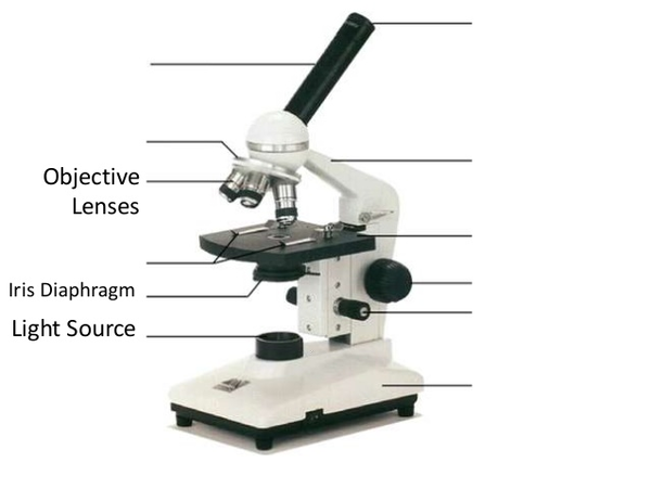
This diaphragm is mostly used to control the contrast. The pathway of light through a compound microscope is. Closing the iris diaphragm will reduce the amount of illumination of the specimen but increases the amount of contrast. Iris diaphragm controls the amount of light reaching the specimen. This diaphragm is located closer to the condenser system of a microscope.
 Source: studylib.net
The diaphragm also called iris is located under the stage and is used to adjust and change the intensity and size of the cone of light that shines up through the side. The general rule is the diaphragm s aperture size is directly proportional to illumination and conversely proportional to contrast while the aperture shape is directly proportional to focus. It is located above the condenser and below the stage. The field iris diaphragm residing in a conjugate plane with the lamp collector lens is imaged sharply into the same plane as the specimen by the microscope condenser. The pathway of light through a compound microscope is.
 Source: blog.microscopeworld.com
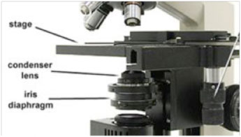
It is located above the condenser and below the stage. In fact the condenser sits right on top of the iris diaphragm. In light microscopy the iris diaphragm controls the size of the opening between the specimen and condenser through which light passes. The diaphragm also called iris is located under the stage and is used to adjust and change the intensity and size of the cone of light that shines up through the side. Iris diaphragm controls the amount of light reaching the specimen.
If you find this site good, please support us by sharing this posts to your favorite social media accounts like Facebook, Instagram and so on or you can also bookmark this blog page with the title microscope iris diaphragm by using Ctrl + D for devices a laptop with a Windows operating system or Command + D for laptops with an Apple operating system. If you use a smartphone, you can also use the drawer menu of the browser you are using. Whether it’s a Windows, Mac, iOS or Android operating system, you will still be able to bookmark this website.


