Pig dissection anatomy
Pig Dissection Anatomy. The incision can be seen in the first photograph below. Follow the steps in the handout to view the external pig anatomy. Securing the pig for the dissection. Fetal pig dissection the pig may or may not be injected with dye.
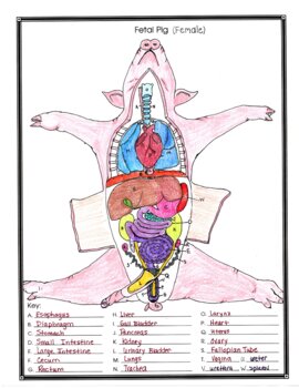 Female Fetal Pig Anatomy And Simulated Dissection Worksheet By Biology Buff From teacherspayteachers.com
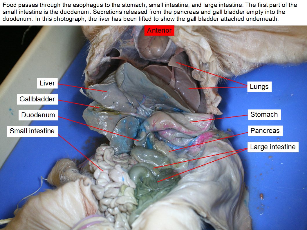
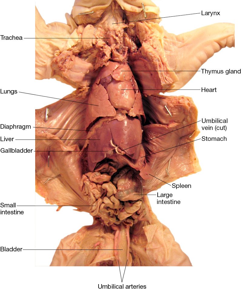
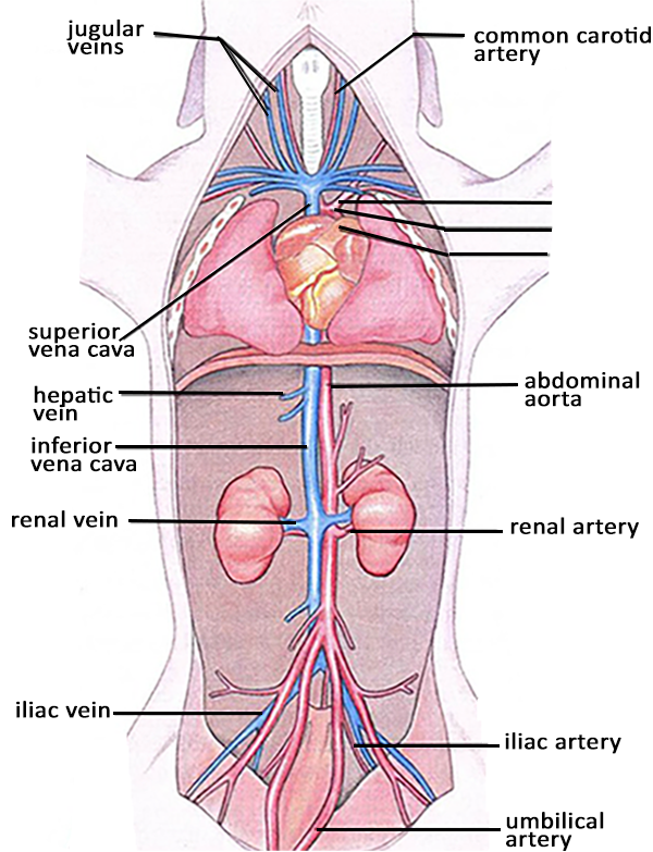
Part 2 includes the remainder of the basic internal anatomy of the pig including the excretory system the lymphatic system the respiratory system and the circulatory system. Part 1 involves external anatomy of the pig and the liver and digestive system. Identify the ureters which carry urine from the kidneys to the urinary bladder find the urinary bladder within the umbillical cord. An incision was made on the side of the neck to enable the injections. Fetal pig dissection the pig may or may not be injected with dye. The arteries have been filled with red latex and the veins with blue.
Access the page reading.
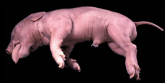
It is fascinating to see how all the organs fit and work together. The incision can be seen in the first photograph below. Mammals are vertebrates having hair on their body and mammary glands to nourish their young. The pigs we are dissecting are called fetal pigs. Fetal pig dissection and fetal pig anatomy fetal pig dissection background. Using the diagram to the left.
 Source: m.youtube.com
The majority are placental mammals in which the developing young or fetus grows inside the female s uterus while attached to a membrane called the placenta. Identify two bean shaped kidneys the dorsal wall in the mid to lower back region the light coloured strip of tissue at the top of each kidney is an adrenal gland. Fetal pigs have not been born. Instead human anatomy can be studied by examining the systems of a pig an animal similar to a human. Continue cutting from the anterior end of this cut so that it resembles an upside down u.

Fetal pig dissection the pig may or may not be injected with dye. An incision was made on the side of the neck to enable the injections. Securing the pig for the dissection. Insert one blade of scissors through the body wall on one side of the umbilical cord and cut posteriorly to the base of the leg as shown in the first photograph below. It is fascinating to see how all the organs fit and work together.
 Source: teacherspayteachers.com
The arteries have been filled with red latex and the veins with blue. Fetal pig dissection and fetal pig anatomy fetal pig dissection background. Instead human anatomy can be studied by examining the systems of a pig an animal similar to a human. The majority are placental mammals in which the developing young or fetus grows inside the female s uterus while attached to a membrane called the placenta. Part 2 includes the remainder of the basic internal anatomy of the pig including the excretory system the lymphatic system the respiratory system and the circulatory system.
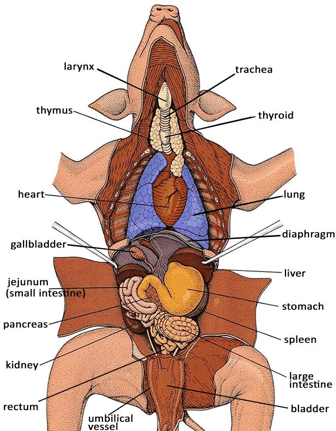 Source: biologycorner.com
Securing the pig for the dissection. Fetal pig dissection and fetal pig anatomy fetal pig dissection background. Using the diagram to the left. Insert one blade of scissors through the body wall on one side of the umbilical cord and cut posteriorly to the base of the leg as shown in the first photograph below. Part 2 includes the remainder of the basic internal anatomy of the pig including the excretory system the lymphatic system the respiratory system and the circulatory system.
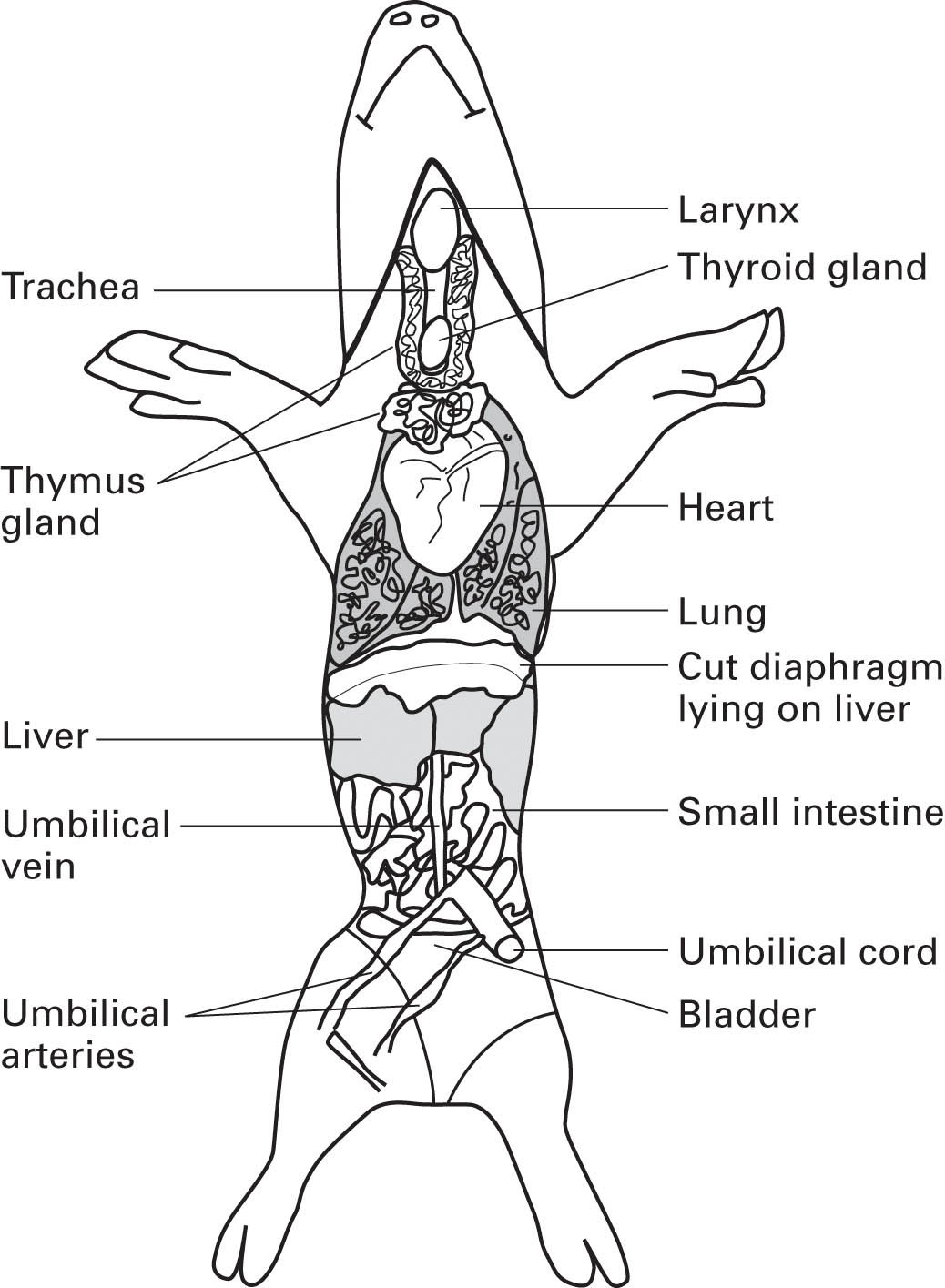 Source: flinnsci.com
The incision can be seen in the first photograph below. An incision was made on the side of the neck to enable the injections. The arteries have been filled with red latex and the veins with blue. Fetal pig dissection and fetal pig anatomy fetal pig dissection background. Fetal pig anatomy dissection sorry guys but you will not get the chance to examine the internal organs of a real human body.
 Source: courses.lumenlearning.com
Insert one blade of scissors through the body wall on one side of the umbilical cord and cut posteriorly to the base of the leg as shown in the first photograph below. Securing the pig for the dissection. Identify the ureters which carry urine from the kidneys to the urinary bladder find the urinary bladder within the umbillical cord. Download a pdf of the lab to print. The majority are placental mammals in which the developing young or fetus grows inside the female s uterus while attached to a membrane called the placenta.
 Source: flinnsci.com
It is also a very exciting dissection because like sheep and their organs the internal anatomy is similar to humans. Part 2 includes the remainder of the basic internal anatomy of the pig including the excretory system the lymphatic system the respiratory system and the circulatory system. This product includes a 2 part fetal pig dissection lab. Continue cutting from the anterior end of this cut so that it resembles an upside down u. The pigs we are dissecting are called fetal pigs.
 Source: whitman.edu
The majority are placental mammals in which the developing young or fetus grows inside the female s uterus while attached to a membrane called the placenta. It is also a very exciting dissection because like sheep and their organs the internal anatomy is similar to humans. Download a pdf of the lab to print. Instead human anatomy can be studied by examining the systems of a pig an animal similar to a human. The arteries have been filled with red latex and the veins with blue.
 Source: pinterest.com
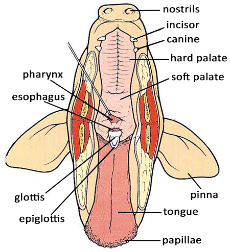
The pigs we are dissecting are called fetal pigs. Follow the steps in the handout to view the external pig anatomy. The fetal pig that you will dissect has been injected with a colored latex rubber compound. The pigs we are dissecting are called fetal pigs. Identify the ureters which carry urine from the kidneys to the urinary bladder find the urinary bladder within the umbillical cord.
 Source: biologycorner.com
The majority are placental mammals in which the developing young or fetus grows inside the female s uterus while attached to a membrane called the placenta. Fetal pig dissection guide project a fetal pig is a great choice for dissection because the size of the organs make them easy to find and identify. Part 1 involves external anatomy of the pig and the liver and digestive system. The fetal pig that you will dissect has been injected with a colored latex rubber compound. It is also a very exciting dissection because like sheep and their organs the internal anatomy is similar to humans.

Access the page reading. Mammals are vertebrates having hair on their body and mammary glands to nourish their young. Follow the steps in the handout to view the external pig anatomy. This product includes a 2 part fetal pig dissection lab. Part 1 involves external anatomy of the pig and the liver and digestive system.
 Source: pinterest.com
Follow the steps in the handout to view the external pig anatomy. The pigs we are dissecting are called fetal pigs. Instead human anatomy can be studied by examining the systems of a pig an animal similar to a human. The fetal pig that you will dissect has been injected with a colored latex rubber compound. Follow the steps in the handout to view the external pig anatomy.
 Source: courses.lumenlearning.com
An incision was made on the side of the neck to enable the injections. The pigs we are dissecting are called fetal pigs. Fetal pig dissection the pig may or may not be injected with dye. Instead human anatomy can be studied by examining the systems of a pig an animal similar to a human. Access the page reading.
 Source: biologycorner.com
Fetal pigs have not been born. Fetal pigs have not been born. An incision was made on the side of the neck to enable the injections. Using the diagram to the left. Identify two bean shaped kidneys the dorsal wall in the mid to lower back region the light coloured strip of tissue at the top of each kidney is an adrenal gland.
 Source: courses.lumenlearning.com
Fetal pig anatomy dissection sorry guys but you will not get the chance to examine the internal organs of a real human body. Securing the pig for the dissection. Identify two bean shaped kidneys the dorsal wall in the mid to lower back region the light coloured strip of tissue at the top of each kidney is an adrenal gland. Mammals are vertebrates having hair on their body and mammary glands to nourish their young. Fetal pig dissection and fetal pig anatomy fetal pig dissection background.
If you find this site helpful, please support us by sharing this posts to your favorite social media accounts like Facebook, Instagram and so on or you can also save this blog page with the title pig dissection anatomy by using Ctrl + D for devices a laptop with a Windows operating system or Command + D for laptops with an Apple operating system. If you use a smartphone, you can also use the drawer menu of the browser you are using. Whether it’s a Windows, Mac, iOS or Android operating system, you will still be able to bookmark this website.


